The result of structure outlining is shown on the VOIS page.
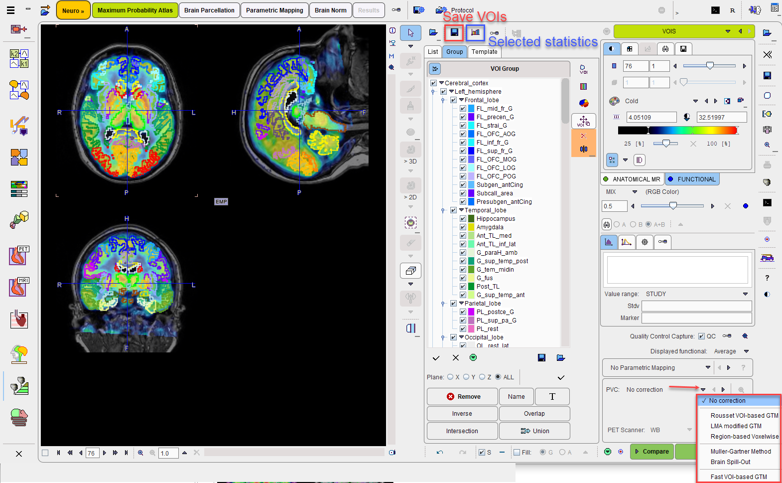
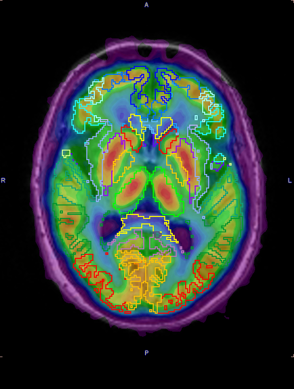
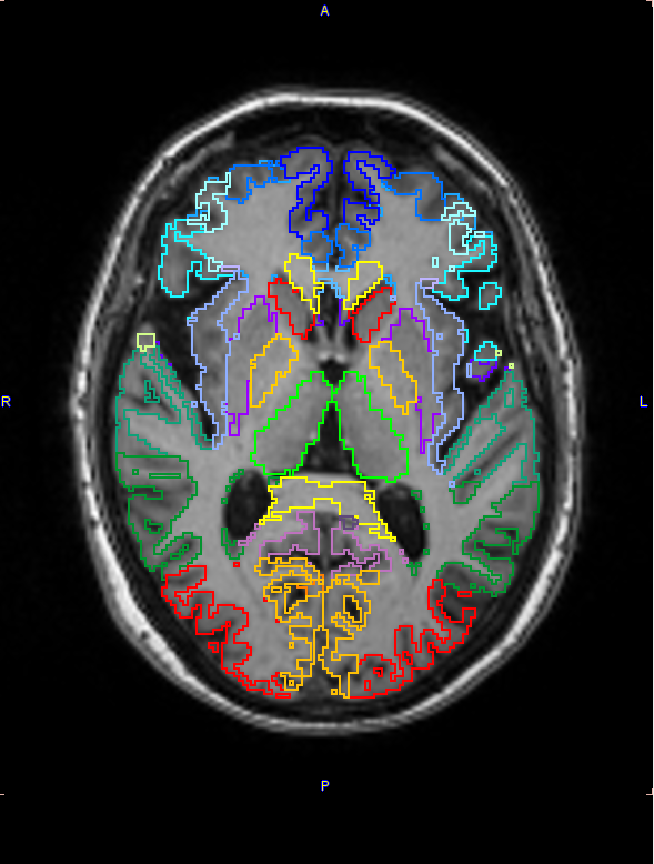
The image display shows a fusion of the MR image with the averaged PET image in the selected Result space. Please use the fusion slider to change the weight between the image contributions, and use the individual image control tabs for the changing the individual image displays. The example below shows the contour VOIs with 50% mixing (on the left) and MR only (to the right).
VOI Editing and Selection
At this time the contour VOIs can be interactively adjusted using the VOI features of PMOD, which are described in the PMOD Base Functionality Guide. Note that the List tab should be selected for the adjustment, and that depending on the configuration only a reduced set of VOI tools may be available.
Please note that for the step by step interactive procedure the regions for which the atlas description is set to H will NOT be selected on the Group tab.
A subset of the VOIs can easily be selected on the Group tab as described above.
Quality Control
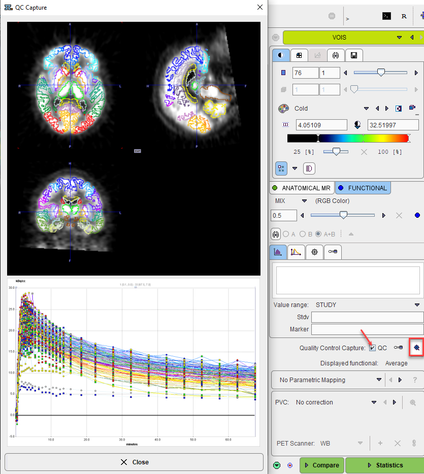
If the QC box is enabled, a screen capture of the VOIs overlayed on the selected functional image will be saved, based on the capture settings as illustrated below:
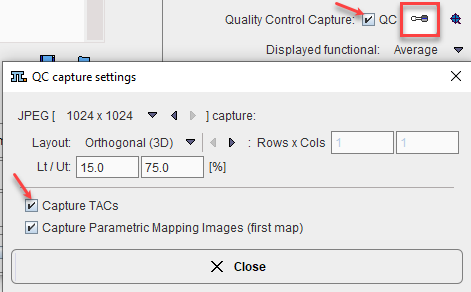
This setting is saved in the protocol and is relevant for batch processing: it allows the user to quickly inspect the outcome of the matching procedures, in the selected result space, at the end of the batch processing.
Partial-Volume Correction (PVC)
The PNEURO tool supports GTM-based partial-volume correction (PVC) of the PET signal. Note that the corrections are sensitive to the signal-to-noise ratio and might produce large artifacts for noisy dynamic data.
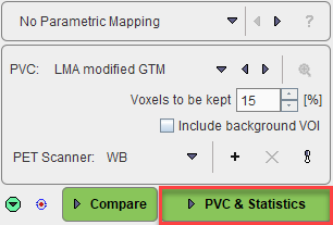
When PVC correction is enabled the right most action button label becomes PVC & Statistics.
The PVC selection has the following choices:
▪No correction: No partial-volume correction is applied (default).
▪Rousset VOI based GTM: The original Rousset GTM correction method is applied.
▪LMA modified GTM: A variant of the Rousset correction method is applied, whereby only a percentage of the pixels in the inner of the VOI is used to calculate the VOI average. This percentage can be set using the Voxels to be kept parameter.
![]()
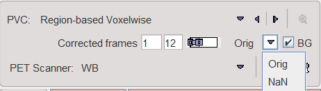
▪Region-based voxelwise: Extension of the GTM method by Thomas et al which performs a voxel-wise correction of the entire image.
▪Muller-Gartner Method: The original Muller-Gartner correction which calculates the corrected activity in the GM pixels by compensating spill-out from the gray matter and spill-in from the white matter.
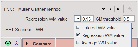
The WM value used for the correction can be specified in 3 ways: as a fix Entered WM value to be entered, as a value determined by linear Regression WM value at WM probability of 1 using the values of all WM pixels, and as the Average WM value in the WM segment above the specified probability threshold.
▪Brain Spill-Out: A variant of the Muller-Gartner correction which compensates only for the spill-out of activity from the brain (WM and GM) to the CSF.
The Brain threshold is used to mask the pixels with brain probability above the specified value.
▪Fast VOI-based GTM: represents actually the LMA modified GTM method which contains some speed-up improvement for the smoothing. The speed performance might be observe when the selected atlas contain a low number of regions.
For the purpose of VOI-based PVC methods, the VOIs complementary to the gray/white matter masked VOIs are taken into account. These VOIs are listed on the Group tab under the Complementar branch once the PVC calculation has been completed.The availability of these VOIs on the list after running the PVC is aimed to increased the transparency such that they can be saved and used for the exact reproduction via the Protocol.
If a PVC method is used, both the original and the corrected statistics are calculated. Note that due to the high number of VOIs the PVC calculation may take several minutes and consumes a significant amount of RAM.
PVC-corrected Images
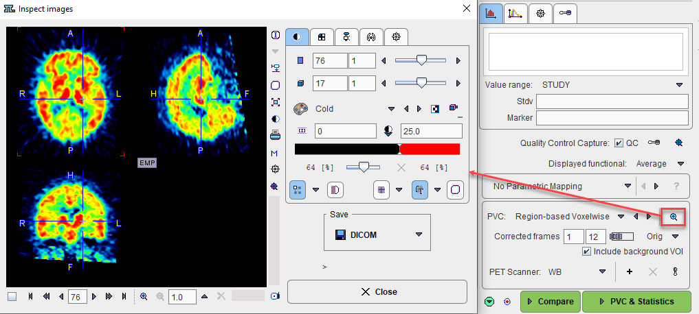
Note that the PVC-corrected images are available for inspection after the PVC&Statistics calculation. When returning to the VOIS page, the inspection button to the right of the PVC method indicated below has become active. It opens the corrected images in a dialog window where they can be interactively inspected and saved.
Statistics Calculation
Once the VOIs have been outlined and carefully checked by the user, it is recommended to first save them, then proceed with calculation of statistics using the Statistics action button.
When PVC correction is enabled the right most action button label becomes PVC & Statistics. The PVC is always calculated on all created VOIs but the statistics values are returned only for the VOIs selected on the Group tab.
The result (for the selected VOIs only) is shown on the separate Result page of the PNEURO tool, from where it can be further evaluated.
Parametric Mapping
If the PET images are dynamic and the PXMOD option is included in the license, parametric mapping using pixel-wise models can be directly applied within PNEURO, as described in a separate section.
When a parametric mapping model is selected the right most action button label becomes Mapping.
Normal Database Comparison
As PNEURO is able to provide the PET images normalized to the atlas space, there is a direct link with the Brain Norm Functionality. The Compare workflow button copies the PET images as they appear on the VOIs page to the Brain Norm Deviation page where they can be compared against the normal uptake pattern for a given tracer, as defined in a normal database (to be defined by the user, see Brain Norm Functionality). Conveniently, if the two spaces match, there is no need to perform a normalization.