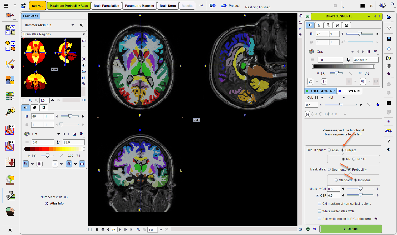
The result of the brain structure transformation is shown on the BRAIN SEGMENTS page. The image on the BRAIN SEGMENTS tab is the transformed atlas with integer labels as the pixel values. It is fused with the MR image.
Result Space
There are three different options to evaluate the PET image, which can be configured using the Result space radio button. The information visualized on the page is updated as soon as the configuration is changed. The image display shows the MR image transformed to the selected result space together with the brain structures.
The Result space options are:
1.Atlas: With this setting the PET image is transformed to the atlas space and the original atlas structures can be applied to it.
2.Subject, MR: With this setting the PET image and the atlas structures are transformed to the MR space.
3.Subject, INPUT: With this setting the atlas structures are mapped to the PET space and statistics are calculated based on the original PET voxel values.
Intersection of the Atlas Structures with Gray Matter
The original atlas structures are typically not restricted to gray matter. Users can take advantage of the gray matter probability calculated during MR segmentation to restrict the structures to pixels with a high probability of belonging to gray matter. This masking is controlled by the elements in the Mask atlas area.
There is a choice between two types of probability information:
•Segments means that the binary information in the segment label image is applied.
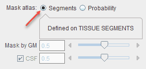
Here, no thresholding is necessary to obtain a GM mask because the binarization was performed at the time of the segment calculation.
•Probability means that a simple threshold is applied to a GM probability map to create a GM mask.
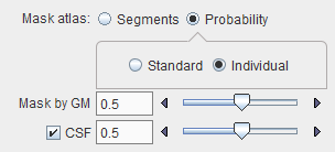
In this case, a Standard probability map can be used which represents a population average, or the Individual GM probability map resulting from the actual MR segmentation. This latter should be the preferred choice unless the segmentation fails.
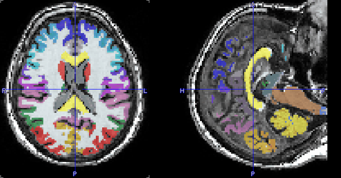
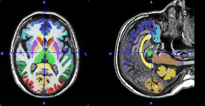
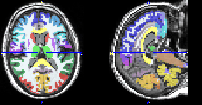
The GM threshold slider of Probability masking allows a lower probability threshold to be defined. This threshold is applied to the GM probability map in order to create a mask. The higher the threshold value, the thinner the cortical structures become. With the CSF box enabled, the CSF threshold slider allows the GM mask to be further trimmed based on voxels with a high probability of being CSF: the higher the threshold the thicker the cortical structures become.
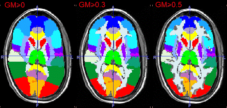
The illustration below shows individual masking at three increasing GM probability thresholds while the CSF box is disabled. The Standard mask will result in broader structures at the same probability threshold.
The illustration below shows individual masking at three decreasing CSF probability thresholds and a constant GM probability threshold of 0.5. The Standard mask results in narrower structures as CSF probability threshold decreases.
 bis")
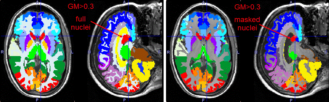
Note that the central structures are not affected by masking in the example above, because the box GM masking non-cortical regions is not enabled. If the option is enabled, the central structures will also be limited based on the grey matter tissue probability mask, as illustrated below. However, because of the low probability levels in that area, the reduction may become too severe. Therefore, masking subcortical regions is disabled by default.
Intersected VOIs and Partial Volume Correction (PVC)
VOI-based PVC correction methods only work properly when all the activity contained in the brain is included in VOIs. Therefore PNEURO automatically creates the full set of complementary VOIs, i.e. GM, WM and CSF fractions of all atlas VOIs, in the memory buffer, regardless of WM masking settings. This full set of VOIs will be used for the PVC calculation, but will not be visible in the user interface and in the result statistics.
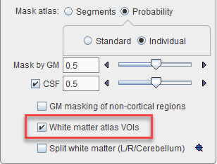
White Matter Atlas VOI
The White matter atlas VOI box, when enabled, allows dividing the white matter region according to the VOIs in the selected atlas.
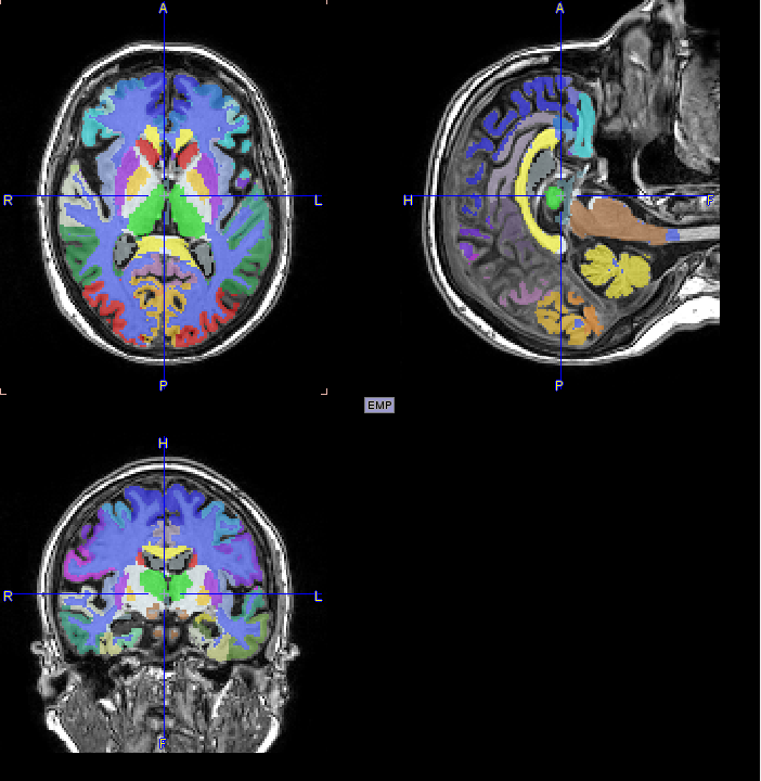
Result on Brain Segments page:
and White_matter VOIs with tree structure:
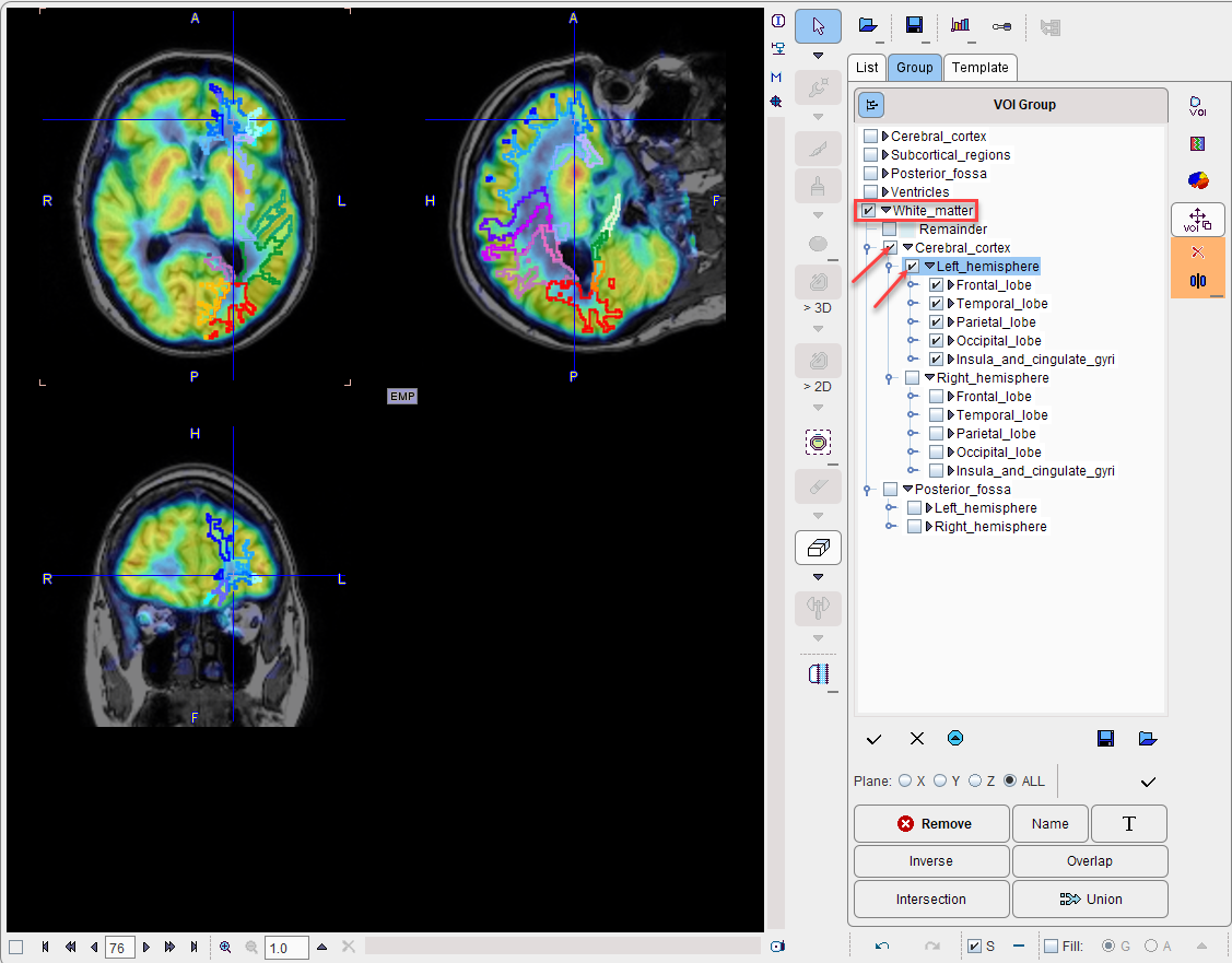
Note: The white matter voxels which do not have any definition in the atlas will be classified as Remainder white matter.
Split Brain
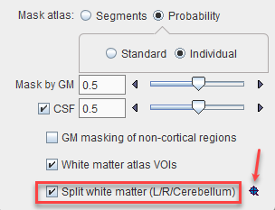
The Split white matter (L/R/ Cerebellum) option is only relevant for the separation of the white matter into left hemispheric, right hemispheric, and cerebellar parts. If the procedure is not enabled or fails, then a global white matter region is created.
Note: The brain split division uses a template in the MNI space. This template consists of three labels corresponding to the left hemisphere, the right hemisphere and the cerebellar part. In the brain split process the brain mask is eroded with an initial erosion size and the separation into individual segments is verified. The result must consist in at least 3 separate segments. If it is not the case the erosion size is incremented with the erosion step. The algorithm verifies each separate region to which label in the template belongs. The front propagation is used to divide the whole brain mask only when all the three region seeds are found to be geodesic.
Inspect the Left/Right Split Outcome
The ![]() Inspect left/right split button opens a dialog window for inspecting the result of the brain splitting procedure.
Inspect left/right split button opens a dialog window for inspecting the result of the brain splitting procedure.
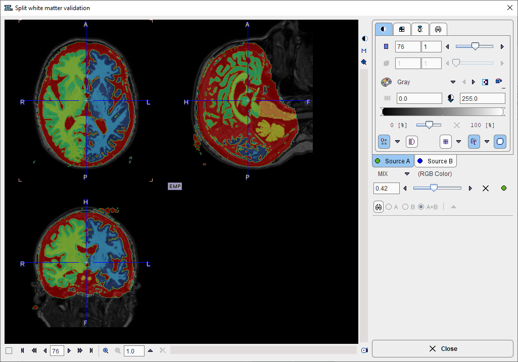
There are 7 resulting segments, which are shown in the Source B tab. They correspond to left white matter, left gray matter , right white matter, right gray matter, cerebellar gray matter (including pons), cerebellar white matter and CSF. If the procedure doesn't come up with a reasonable splitting into right and left parts, it is recommended to Close the window and switch OFF the Split check box.
Structure Outlining
Once the result space and gray matter masking have been specified, the brain structures are fully defined and can be outlined to create contour VOIs. This process is started with the Outline action button.