Image normalization applied for [11C]PK11195 [8] and [11C]PIB [9] consists of a z-score calculation, which can be performed in the PMOD viewing tool (PVIEW).
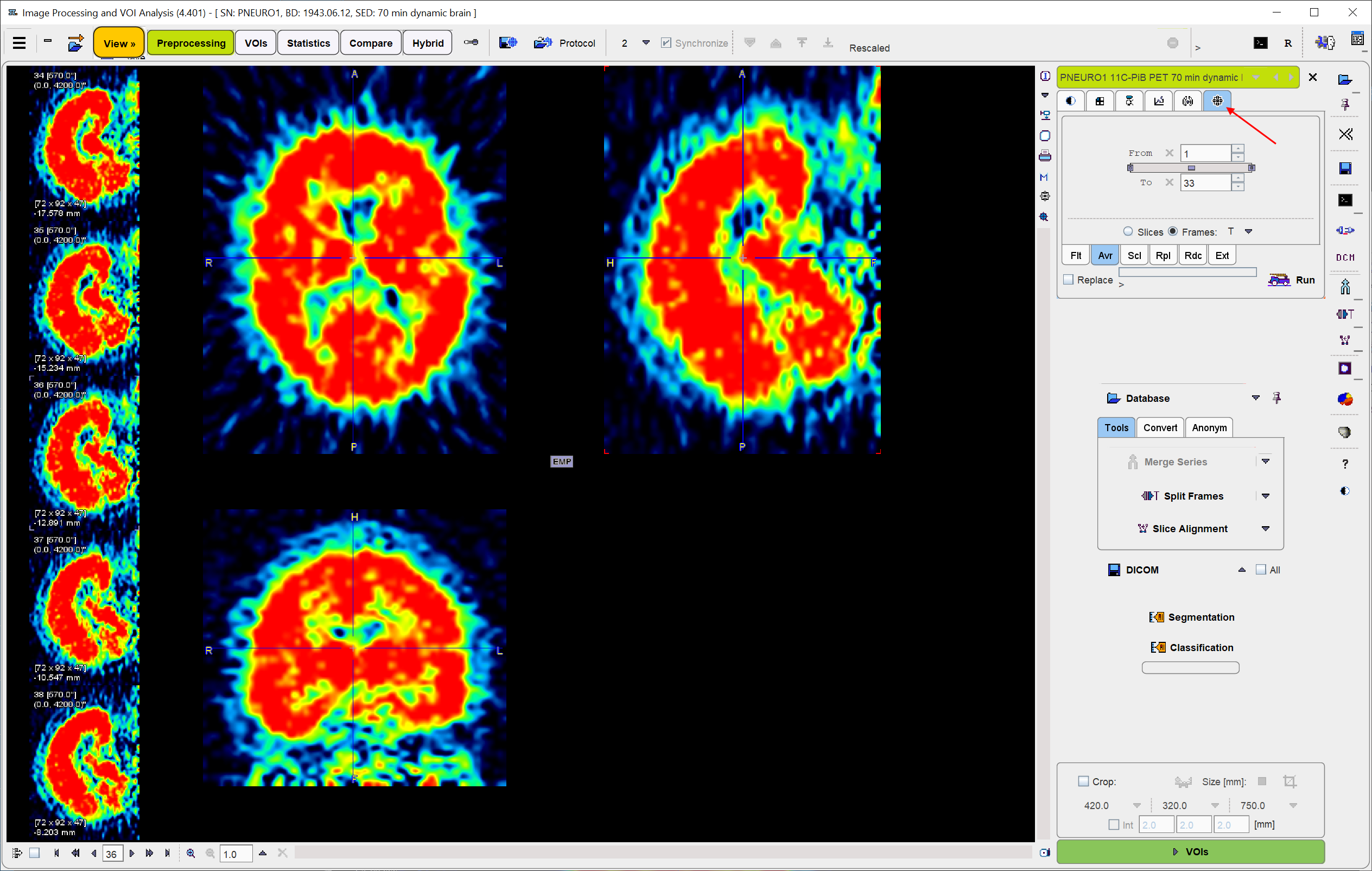
In PVIEW, first load the dynamic PET data. Next, average the image frames to get a better anatomical image.
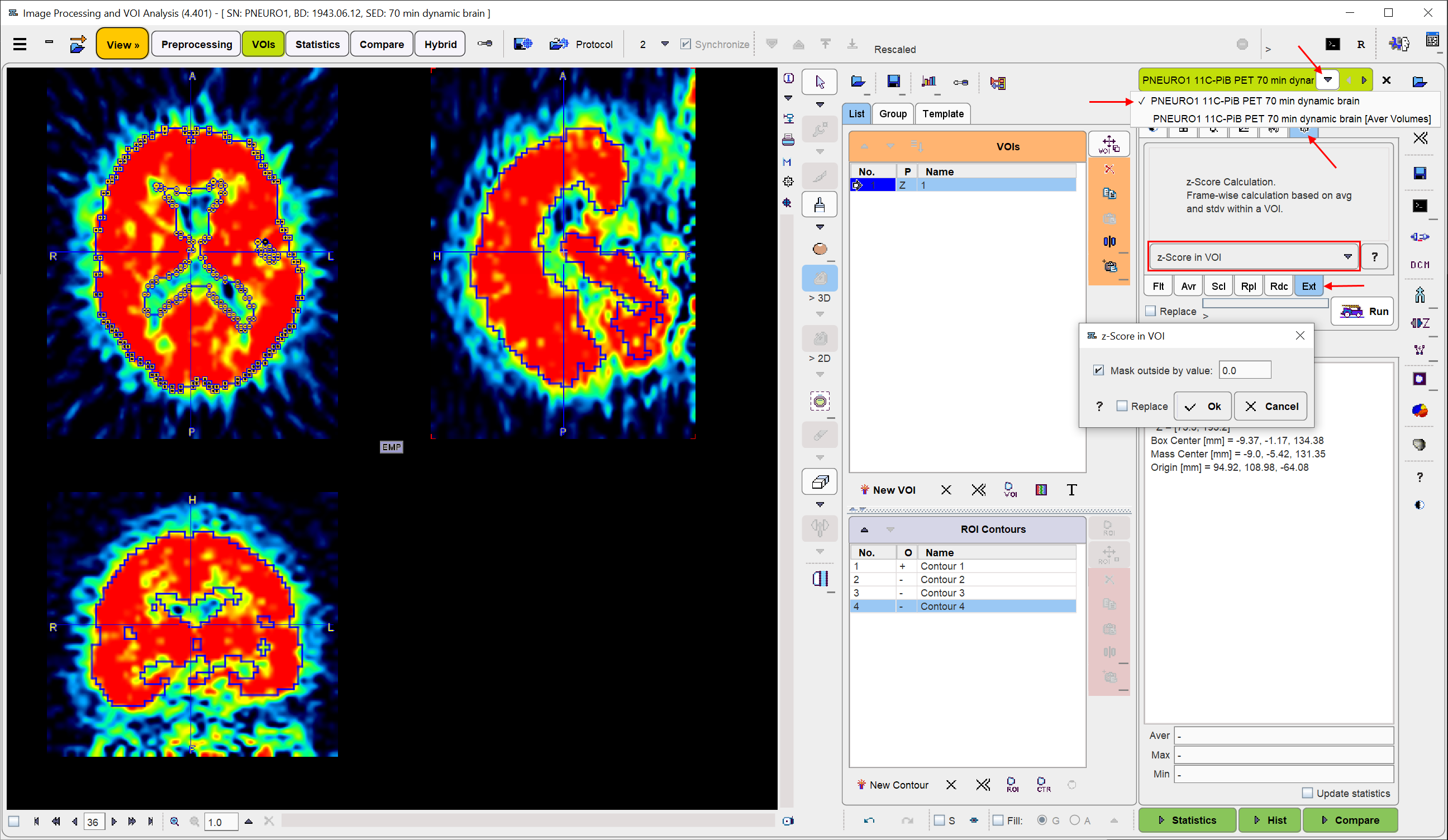
Use the averaged image for defining a bounding VOI for the mean and standard deviation calculation. In the example below the VOI page was selected, the 3D iso-contouring tool activated, and by clicking at the brain edge a VOI obtained which encloses the brain. Alternatively, other approaches can be developed which define the VOI more objectively. Ikoma et al. [9] used a VOI enclosing grey and white matter.

After VOI creation, switch back to the original data, select the tools tab, the Ext sub-tab, and start z-score calculation with the Run button.
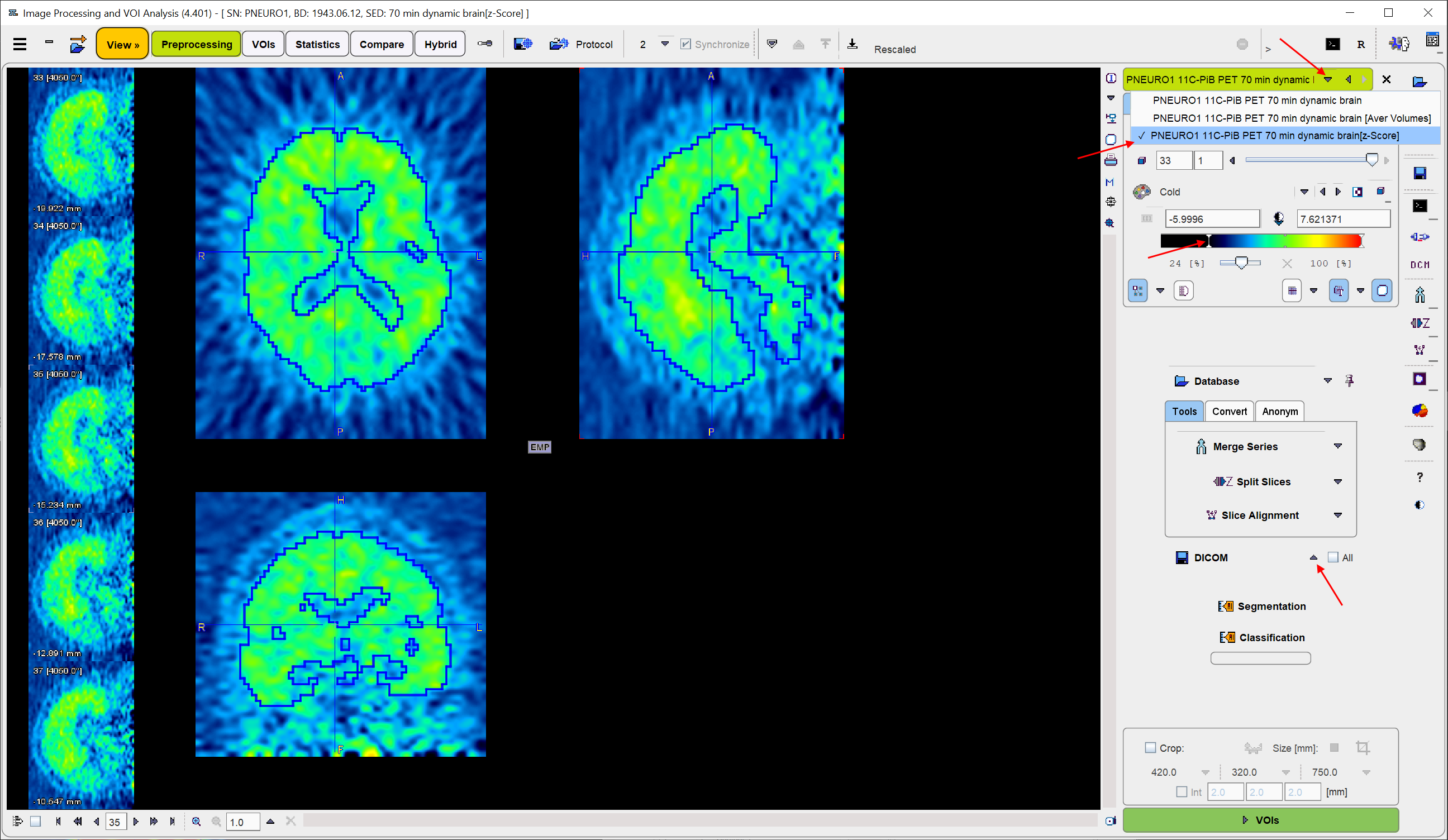
The resulting normalized dynamic images are added as the third series. It can be saved on the View page as illustrated below. Note that the color table thresholds may not be adequate any more due to the change of the value range, and may need adjustments.