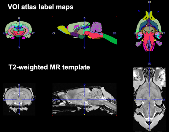
The Rat (Waxholm-80um) atlas is based on the brain of a 397.6 g male Sprague-Dawley rat. The brain was imaged with MR ex vivo, resulting in T2-weighted gradient echo images with 40 um isotropic spatial resolution. 76 brain structures were segmented [1, 2].
E. A. Papp prepared a version suitable for PMOD and made it available for download on nitrc.org: https://www.nitrc.org/projects/whs-sd-atlas/
An adapted version of this atlas is distributed with PMOD as a convenience to our users. Because the original 40um isotropic resolution is beyond typical in vivo MR and far beyond the resolution applicable for PET image analysis, it was down-sampled to 80um. The original text file associated with the atlas was re-organized to consecutively number the regions. Note: down-sampling left no actual voxels in regions 6,33,47,49,60,63 as numbered in the atlas text file.
Atlas Label Map and MR Normalization Template
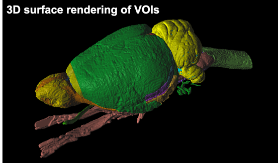
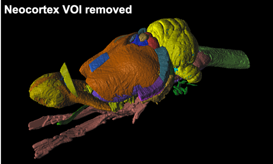
3D Rendering of Atlas Regions
References
[1] Papp EA, Leergaard TB, Calabrese E, Johnson GA, JG (2014). Waxholm Space atlas of the Sprague Dawley rat brain. NeuroImage 97:374-386. doi: 10.1016/j.neuroimage.2014.04.001
[2] Kjonigsen LJ, Lillehaug S, Bjaalie JG, Witter MP, Leergaard TB (2015). Waxholm Space atlas of the rat brain hippocampal region: Three-dimensional delineations based on magnetic resonance and diffusion tensor imaging. NeuroImage 108:441-449. doi: 10.1016/j.neuroimage.2014.12.080