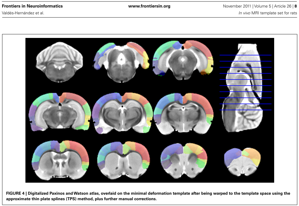
Overview
The Wistar Rat (Tohoku) atlas was developed by Valdes-Hernandez et al [1] using 7T T2-MRIs from 30 Wistar rats. The template image was constructed as a "minimal-deformation" template, and the coordinates are thus not in Paxinos space. In the same space, gray matter, white matter and CSF probability maps were calculated, so that the 3-probability maps normalization can be applied in addition to the template-based normalization. 48 bi-lateral cortical structures were digitized from the Paxinos atlas and registered them to the template image. This atlas can be used for VOI statistics and for the anatomical interpretation of fMRI results.
The atlas is distributed with PMOD by courtesy of Prof. Akira Sumiyoshi, Tohoku University, Japan (atlas website).
Spatial Normalization
Two normalization approaches are supported:
▪Template-based normalization using the Wistar Rat (Tohoku)-MR skull-stripped T2-MR template illustrated above.
▪3-tissue probability maps normalization using Gray matter, White matter and CSF combined in a single tpm.nii file.
The images of these templates can be found in the resources/templates/voitemplates/Wistar Rat (Tohoku)/normalization directory.
Reference
[1] Valdes-Hernandez PA, Sumiyoshi A, Nonaka H, Haga R, Aubert-Vasquez E, Ogawa T, Iturria-Medina Y, Riera JJ, Kawashima R: An in vivo MRI Template Set for Morphometry, Tissue Segmentation, and fMRI Localization in Rats. Frontiers in neuroinformatics 2011, 5:26. DOI