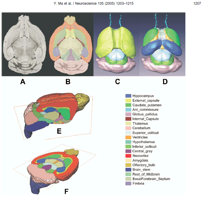
For the analysis of mouse brain data the Mouse (Ma-Benveniste-Mirrione) VOI atlas [1,2] is available. It represents the minimum deformation atlas of 10 C57BL/6J mice (male, 12-14 weeks, 25-30g). We would like to thank Helene Benveniste and Martine Mirrione for providing the data and helping with the integrations.
Spatial Normalization
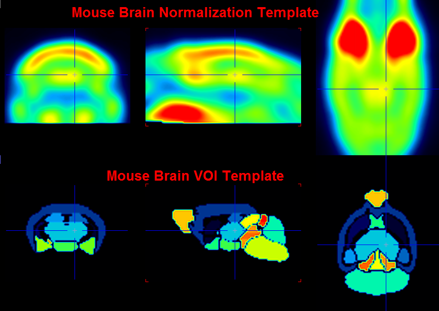
Two normalization templates are available in the fusion tool:
▪Mouse (Ma-Benveniste-Mirrione)-FDG: This is an FDG baseline PET template as illustrated below.
▪Mouse (Ma-Benveniste-Mirrione)-T2: This is a T2-weighted MRI PET template which is in the same space as the PET templates.
The images of these templates can be found in the resources/templates/voitemplates/Mouse (Ma-Benveniste-Mirrione)/normalization directory.
VOI Atlas
The VOI atlas Mouse (Ma-Benveniste-Mirrione) can be selected in the list of included template VOIs. The corresponding files can be found in the resources/templates/voitemplates/Mouse (Ma-Benveniste-Mirrione) directory.
References
[1] Ma Y, Hof PR, Grant SC, Blackband SJ, Bennett R, Slatest L, McGuigan MD, Benveniste H. A three-dimensional digital atlas database of the adult C57BL/6J mouse brain by magnetic resonance microscopy. Neuroscience. 2005;135(4):1203-15. DOI
[2] Mirrione MM, Schiffer WK, Fowler JS, Alexoff DL, Dewey SL, Tsirka SE. A novel approach for imaging brain-behavior relationships in mice reveals unexpected metabolic patterns during seizures in the absence of tissue plasminogen activator. Neuroimage. 2007;38(1):34-42. DOI