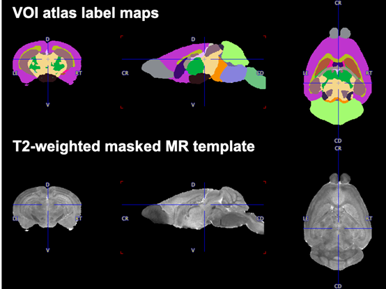
The Mouse Brain (Waxholm-40um) atlas represents the canonical Waxholm Space (WHS) adult C57BL/6J mouse brain. It is based on histology and high spatial resolution MR of adult male C57BL/6J mice aged 66-78 days [1]. The atlas resources are available at https://www.nitrc.org/projects/incfwhsmouse.
An adapted version of this atlas is distributed with PMOD as a convenience to our users. Because the original 21.5um isotropic resolution is beyond typical in vivo MR and far beyond the resolution applicable for PET image analysis, it was down-sampled to 40um. It provides a higher resolution space than the Mouse (Ma-Benveniste-Mirrione) atlas. PET templates corresponding to the mouse Waxholm space have been reported in the literature [2].
The Mouse Brain (Waxholm-40um) atlas contains 26 structures (+ inner ear) organized in accordance with the NeuroLex Brain Partonomy scheme. An ex-vivo masked T2-weighted MR template image is provided for normalization.
Atlas Label Map and Normalization Template
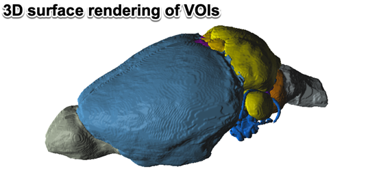
3D Rendering of Brain Regions
References
[1] G. Allan Johnson, Alexandra Badea, Jeffrey Brandenburg, Gary Cofer, Boma Fubara, Song Liu, Jonathan Nissanov. Neuroimage 53 (2) 365-372, 2010. PMCID: PMC2930145.
[2] Bertoglio D, Verhaeghe J, Miranda A, Kertesz I, Cybulska K, Korat Š, Wyffels L, Stroobants S, Mrzljak L, Dominguez C, Liu L, Skinbjerg M, Munoz-Sanjuan I, Staelens S. Validation and noninvasive kinetic modeling of [11C]UCB-J PET imaging in mice. J Cereb Blood Flow Metab. 2020 40(6):1351-1362. doi: 10.1177/0271678X19864081.