Overview
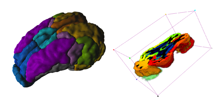
The Pig (CH.Malbert) atlas is based on brain scans of 5 6-month-old female domestic pigs (Sus scrofa domesticus). An ex vivo T2-weighted MRI was used by Saikali et al. [1] to construct the atlas, and a T1 MRI is provided for image coregistration. Subsequently, the same group constructed a 99mTc-HMPAO SPECT template [2] and FDG PET template [3] in the same image space. Additionally, an FDG PET template for Yucatan miniature pigs was developed in the same image space as the domestic pig atlas [4]. The atlas was initially constructed at high resolution (100 x 150 x 100 um) in one hemisphere, and then mirrored to create the full brain atlas. The VOI atlas contains 173 cortical and subcortical regions (reduced from 178 in the original high resolution atlas). The atlas is distributed with PMOD by courtesy of Dr. Charles-Henri Malbert, director of research at the Unité Ani-Scan, Département de Nutrition Humaine, INRA, France.
Spatial Normalization
Four normalization templates are available in the Fuse It tool. They can be selected on the Reference page using the Template button when Pig is selected as Species:
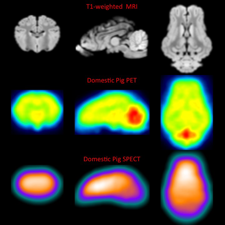
▪Pig (CH.Malbert)-FDG: This is an FDG PET template derived from static scans in 4 month old domestic pigs, shown below.
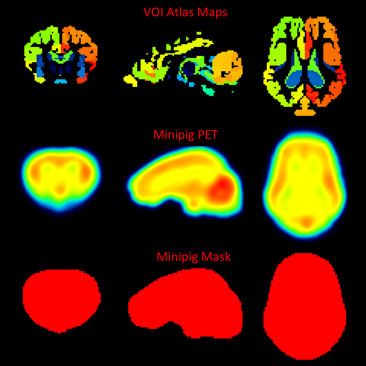
▪Pig 2y (CH.Malbert)-FDG: This is an FDG PET template derived from dynamic scans in 2 year old minipigs, shown below. Correspondence to the domestic pig template and MRI is high.
▪Pig (CH.Malbert)-SPECT: This is a 99mTc-HMPAO SPECT template derived from scans in 4 month old domestic pigs.
▪Pig (CH.Malbert)-T1: This is a T1-weighted MRI PET template in the same image space as the PET templates. The VOI atlas was derived from this image.
The image files corresponding to these templates can be found in the resources/templates/voitemplates/Pig (CH.Malbert)/normalization folder. Additionally, mask files corresponding to each template, and tissue probability maps are available.
Selection and generation of appropriate masks is critical to successful spatial normalization with these templates. The mask corresponding to the template selected should be identified in the Normalization Settings dialog. The PBAS functionality (e.g. via the View tool) can be used to generate a mask for the input image. Accurate exclusion of activity outside the brain, and the olfactory bulbs, is necessary. This user-defined mask should also be identified in the Normalization Settings dialog.
VOI Atlas
The VOI atlas Pig (CH.Malbert) can be selected in the list of included VOI atlases. The corresponding map files in Nifti format can be found in the resources/templates/voitemplates/Pig (CH.Malbert) directory. The brain VOIs are structurally organized in a tree on the Group tab of the VOI editing page. The selection of a VOI subset is supported by a dedicated user interface.
References
1.Saikali S, Meurice P, Sauleau P, Eliat PA, Bellaud P, Randuineau G, Vérin M, Malbert CH. A three-dimensional digital segmented and deformable brain atlas of the domestic pig. J Neurosci Methods. 2010; 192(1):102-9. DOI link: http://dx.doi.org/10.1016/j.jneumeth.2010.07.041.
2.Boubaker J, Val-Laillet D, Guérin S, Malbert CH. Brain processing of duodenal and portal glucose sensing. J Neuroendocrinol. 2012 Aug;24(8):1096-105. DOI.
3.Gaultier A, Meunier-Salaün MC, Malbert CH, Val-Laillet D. Flavour exposures after conditioned aversion or preference trigger different brain processes in anaesthetised pigs. Eur J Neurosci. 2011 Nov;34(9):1500-11. DOI.
4.Ochoa M, Malbert CH, Meurice P, Val-Laillet D. Effects of Chronic Consumption of Sugar-Enriched Diets on Brain Metabolism and Insulin Sensitivity in Adult Yucatan Minipigs. PLoS One. 2016 Sep 1;11(9):e0161228. DOI.