The active Epi/Endo segmentation relies on manual segmentation of the boundaries of endocardium, epicardium and optionally right ventricle. This approach is always a resort if the automatic procedures don't result in a useful segmentation.
Preprocessing
Please follow these steps:
1.Switch segmentation to Epi/Endo Segmentation.
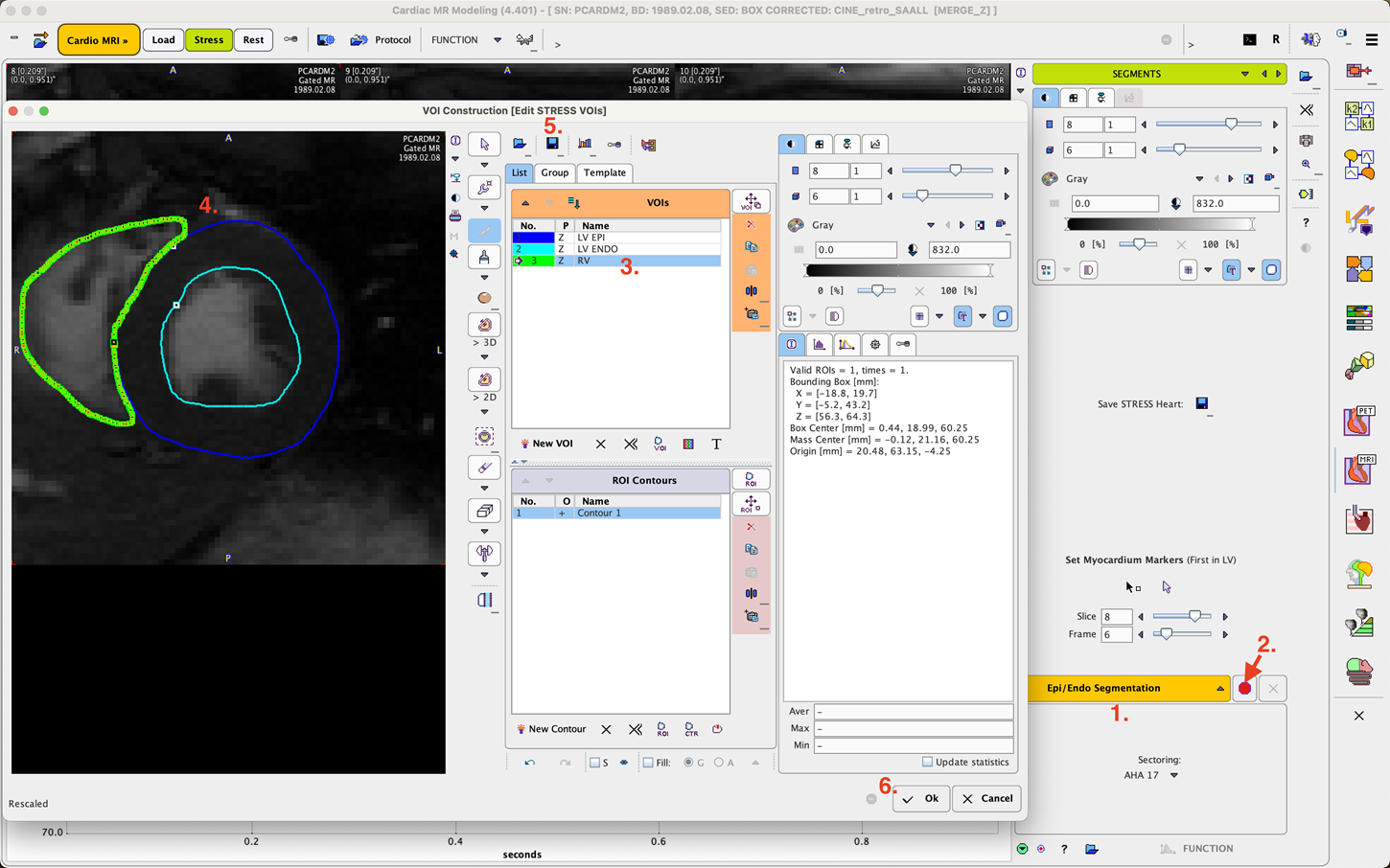
2.Activate the red bullet button to open the VOI Construction dialog window illustrated below.
3.Select a segment in the VOIs list, and define the corresponding boundary contours on all slices in all frames of the image series. In the case of a clear contrast iso-contouring can efficiently trace the LV and RV boundaries as in the example shown. For the epicardium, manual might be required. Different tools are available to work most efficiently. Please refer to the related section in
4.Click into the center of the myocardial muscle at the location, where the RV ends
5.Save the contour VOIs to be able to get back to easily do adjustments. Note: if a protocol is saved, the current VOIs will be included and loaded when restoring the protocol.
6.Close the window with Ok to return to the SEGMENTS page.
Now function can be analyzed as described below.