The twilite measures the radioactivity in a catheter which runs between two LYSO/BGO crystals. In animals a very efficient solution is to install a shunt between the femoral artery and the femoral vein. This allows continuous measurement of the whole blood activity without any loss of blood. Such arteriovenous shunts can be placed in animals as small as a mouse, using catheters of an appropriate size (e.g. mouse: PE10, rat: PE50). A typical setup and schematic are shown below.
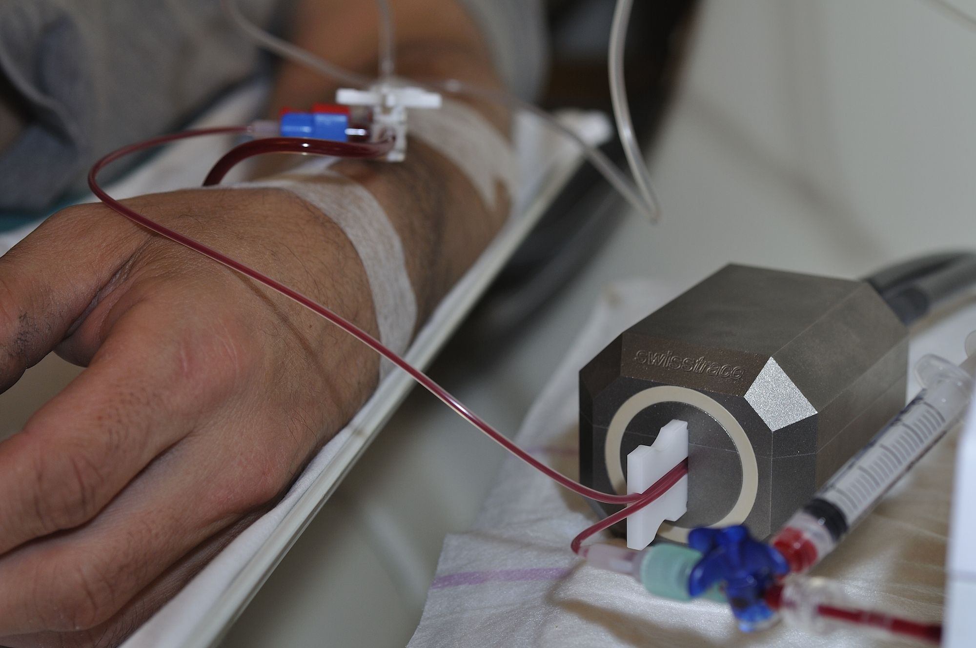
In humans and larger animals, the withdrawn blood should not flow back into the body. One usually places a catheter into the radial artery, runs the catheter through the twilite measuring head and directs the blood into a waste container. A schematic is shown below.
In all cases, swisstrace recommends that the blood flow in the catheter is controlled by a suitable pump. However, in small animals, it is also possible to let the shunt run freely driven only by the arteriovenous pressure difference.
The appropriate catheter diameter differs according to the size of the animals. Swisstrace delivers templates to guide the catheter through the measuring head for each catheter size, such that a well-defined geometry is established. The characteristics of the shunt are described in a paper published by Weber and co-workers [1], and further information on shunt and general twilite setup can be found in [2] and [3].
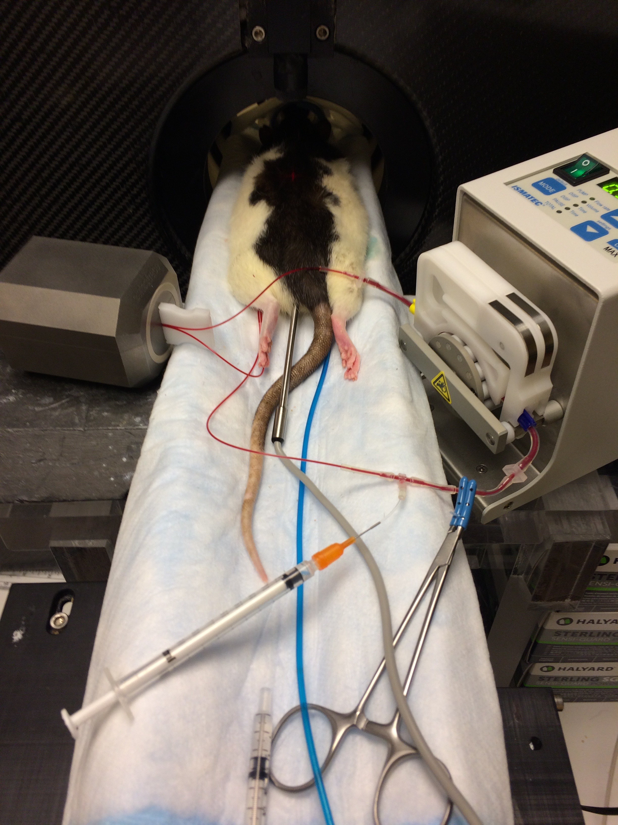
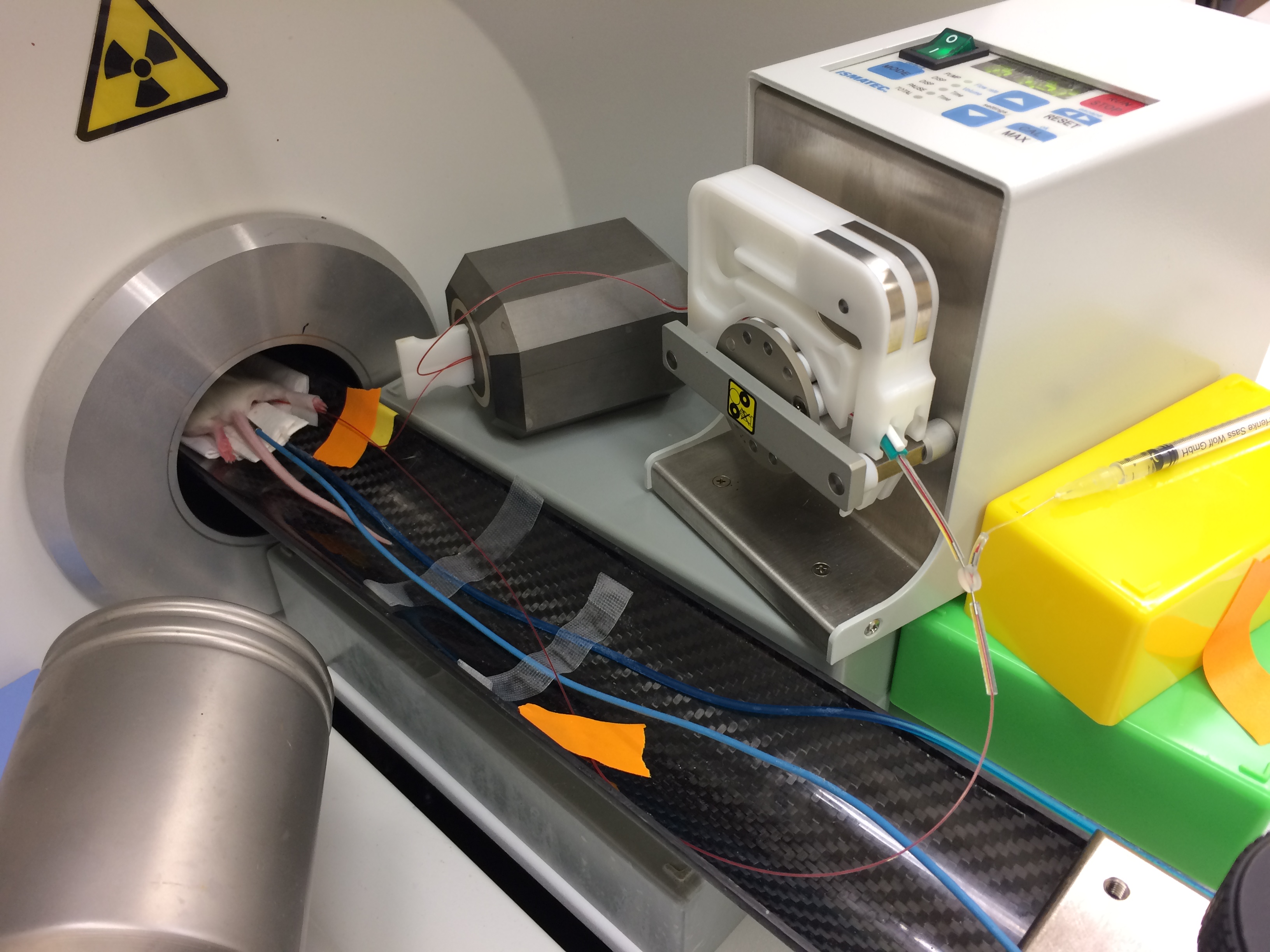
A typical setup for a quantitative rat PET experiment is illustrated below:

1.Shunt running from the femoral artery to the femoral vein.
2.Peristaltic pump to control blood flow in the shunt.
3.Twilite measuring head made of tungsten.
4.Light guides carrying the photons from the crystals to the PMT‘s. These guides have a standard length of 2 m; they can be as long as 16 m (8 + 8 m) in MR compatible systems.
5.Base unit with coincidence electronics (twilite two shown).
6.Touch screen for device setup and start/stop.
7.Not seen here: TCP/IP connection on rear panel of data acquisition box, computer with PMOD PSAMPLE data acquisition and data analysis software.
Schematic for Rats and Mice

Shunt functions
1.Catheter running femoral artery to the femoral vein.
2.Peristaltic pump.
3.Twilite measuring head made of tungsten.
4.LYSO crystal 1.
5.LYSO crystal2.
6.Light guides carrying the photons from the crystals to the PMT‘s. These guides have a standard length of 2 m, they can be as long as 16 m (8 + 8 m) in MR compatible systems.
7.Base unit with coincidence electronics.
8.TCP/IP connection to computer with PMOD data acquisition tool PSAMPLE.
9.Computer with PMOD data acquisition and analysis.
The arteriovenous shunt can serve several additional functions, such as blood pressure monitoring, tracer injection, as well as the collection of blood samples for metabolite analysis. The procedure illustrated below is recommended for collection of blood samples: a small cut is made into the catheter using a scalpel. In normal operation the catheter is bent upwards, so that the cut is closed and blood circulates. To obtain blood samples the shunt is briefly pressed downwards in order to open the cut and blood drops can be collected with minimal dead volume.
Additional detail of the experimental setup is seen here:
Rat
Mouse
Schematic for Humans
In humans there is no return of arterial blood.

1.Catheter running from the radial artery to the waste container.
2.Twilite measuring head made of tungsten.
3.LYSO crystal 1.
4.LYSO crystal 2.
5.Light guides carrying the photons from the crystals to the PMT‘s. These guides have a standard length of 2 m, they can be as long as 16 m (8 + 8 m) in MR compatible systems.
6.Base unit with coincidence electronics.
7.TCP/IP connection to computer with PMOD data acquisition tool PSAMPLE.
8.Computer with PMOD data acquisition and analysis.
9.Peristaltic pump.
10.Waste container.
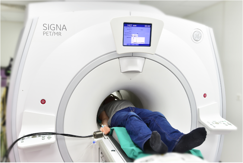
A typical configuration for PET/CT is seen here:
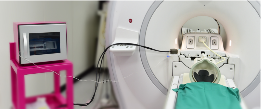
And a typical configuration for PET/MR is seen here: