The sequential steps for converting blood measurements to an input curve are explained in a dedicated section. The current plasma fraction model serves for converting a whole-blood activity curve into a plasma-activity curve.
It is assumed that a curve representing the measured fractions of plasma/whole-blood activity has been loaded with Load Plasma/WB Fraction.
Operational Model Curve
The 3 Exponentials model allows replacing the tail of the fraction curve by a fitted sum of up to three exponentials. The Begin parameter defines at what time the models switches from linear to tri-exponential interpolation. The exponentials are each defined by an Amplitude and a Halftime [min] of the decay. The number of exponentials can easily be reduced by fixing one or two of the amplitudes at a value of 0.
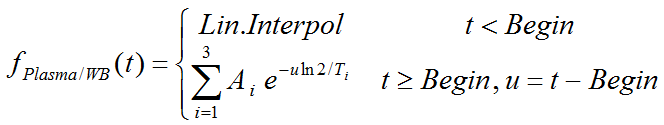
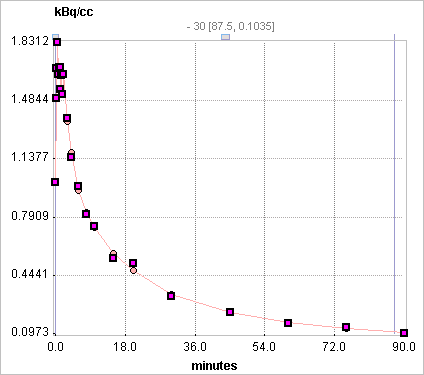
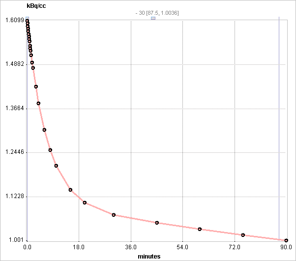
The illustration below shows an example with the exponentials fitted following 2 minutes (Begin = 120sec). Note that the linear interpolation assumes a fraction of 1 at t=0.
Parameter Fitting
The model supports the fitting of the parameters Begin, Amplitude 1, 2, 3 (=A1, A2, A3) and Halftime 1, 2, 3 (=T1, T2, T3). Fitting is started with the Fit plasma fraction button, which only takes the samples after the Begin time into consideration.
Appropriate default parameters for FDG scans:
▪Rat experiments [1]: Plasma/whole_blood(t) = 0.51 e(-ln2/4.79 t)+0.3 e(-ln2/337 t) +0.8
▪Mouse Experiments [2]: Plasma/whole_blood(t) = 0.386*e(-0.191*t)+ 1.165
Use without Measured Fraction
If no measured plasma/whole-blood fraction data is available, the 3 Exponentials model can still be applied. In this case the Begin time is disregarded, and the functional defined by the entered parameters applied.
References
1.Weber B, Burger C, Biro P, Buck A: A femoral arteriovenous shunt facilitates arterial whole blood sampling in animals. Eur J Nucl Med Mol Imaging 2002, 29(3):319-323.
2.Ferl GZ, Zhang X, Wu HM, Kreissl MC, Huang SC: Estimation of the 18F-FDG input function in mice by use of dynamic small-animal PET and minimal blood sample data. J Nucl Med 2007, 48(12):2037-2045. DOI