Findings by Rousset et al. [2]
According to Rousset et al. [2], the accuracy of the GTM method depends primarily on the proper identification of the tissues which have different functional properties. If this is the case, the GTM algorithm is capable of accurately correcting the regional concentration within small structures such as the human basal ganglia. Furthermore, the propagation of statistical noise during partial-volume correction was found to be easily predictable and suitable for the application in dynamic PET.
Findings with the PMOD Implementation
The application of the GTM method to simulated PET images with realistic, spatially variant PSF resulted in the following observations (unpublished work Olivier Barret, PhD, Institute for Neurodegenerative Disorders, New Haven, CT, USA):
▪GM VOIs are always corrected into the right direction, up- and downwards.
▪A FWHM of 7mm is a reasonable default setting for the Gaussian PSF.
▪If a subject MRI is available, the individual segments result in a better partial-volume correction than the standard GM, WM and CSF segments.
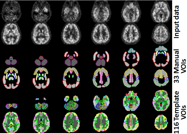
Shown below is an example of a subject data set corrected with 33 manually outlined VOIs, and with 116 VOIs derived from the AAL template combined with individual segments. Note the homogeneous values within the VOIs which are equal the partial-volume corrected average value of each VOI.