PET and SPECT images are inherently affected by the partial-volume effect. This term means that the measured tracer activity concentrations are not accurate due to the relatively low image resolution and the limited tissue sampling. Basically, the low resolution causes a blurring of the image, so that high activities (from a hot lesion) are spread to the surrounding as illustrated below. This effect is called spill-out. The same effect also causes a spill-in of background activity into the volume of interest.
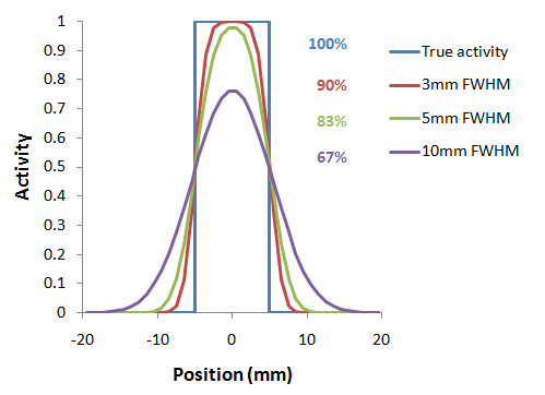
As a consequence, hot lesions tend to appear less aggressive (reduced maximum) but bigger (spreading) than they are in reality.
Spill-in and spill-out depend on the geometry of the objects, the activity distribution of the tracer, and on the resolution of the scanner which may vary across the imaging field-of-view. Therefore, practical correction approaches have to assume certain conditions and can only be approximate. For a nice overview of the topic please refer to the publication of Soret et al. [1].
Solutions Implemented in PMOD
PMOD provides two PVC solutions:
1.PVC VOI-based: This correction is based on the assumption, that the imaging volume can be separated into tissue volumes (VOIs) with homogeneous uptake. If the resolution of the PET scanner is known, the mutual signal contaminations across the VOIs can be calculated and corrected for. This method is known as the GTM (Geometric Transfer Matrix) method and was introduced by Rousset et al. [2,3].
The implementation in PMOD allows the user applying the GTM correction with any set of manually outlined VOIs. Additionally, for the analysis of human brain uptake, the user may take advantage of standard VOIs which are automatically adjusted to the subject's anatomy.
2.PVC Brain MR based: This correction is based on the assumption that white matter uptake is homogeneous. All brain pixels are classified as white matter (WM) or grey matter (GM) and sorted into respective segments. Based on these segments and the assumed PET resolution the spill-out from WM to GM can be estimated and subtracted. Similarly, the spill-out from GM to the surroundings can be estimated and compensated for. The result is a grey matter image with corrected activity values in all pixels. This method was introduced by Muller-Gartner et al. [4].
Given a brain PET and an anatomical MRI of a subject, the implementation in PMOD allows the user performing the segmentation and apply the Muller-Gartner PVC in a fully automated way.
The two PVC methods are implemented as external tools, although they have a much broader scope and require more user interaction than typical external tools. Therefore, their implementations are described in the respective sub-sections below.
Step-wise and Background Calculation Modes
The different PVC methods typically involve several processing steps, some of them using input parameters. There are two ways how a user can apply the PVC calculation:
1.Background calculation: The user enters all required information and adjusts the different parameters, and then starts the processing. The tool window closes, the computations run in a separate thread in the background, and finally returns the corrected images for statistical analysis.
2.Step-wise processing: The user performs the processing steps one by one interactively with a ![]() button, and inspects the intermediate results in the tool. If required, he can change a parameter and initiates the calculation again. After the last step, the final results are returned and the tool closed.
button, and inspects the intermediate results in the tool. If required, he can change a parameter and initiates the calculation again. After the last step, the final results are returned and the tool closed.
The step-wise processing mode has the advantage that all intermediate data remains available, and processing can be repeated from any intermediate step. When data is returned, it is already quality checked, whereas in the background mode the user can not always be sure that all processing steps were successful without inspecting some of the supplementary images returned by the tool.
The All Steps button performs all processing steps sequentially, but does not close the dialog window so that after a first run the different steps can be interactively explored.
Note that the parameters can always be reset to the recommended default values by the ![]() button to establish a well defined situation after some experiments.
button to establish a well defined situation after some experiments.
Common Requirements for Partial-volume Correction in PMOD
The PVC methods implemented in PMOD assume a homogeneous, Gaussian-shaped point-spread function (PSF) of the scanner which is specified by its full-width at half maximum (FWHM) in all directions. The user needs to determine reasonable FWHM values for his reconstructed images and specify them to the algorithm. If possible, a PET reconstruction method with homogeneous resolution should be used, so that the assumption of a stationary PSF is justified. Furthermore, the brain PET images should be reconstructed with a small pixel size not larger than 2.5 mm.
Important Note: The PVC tools expect that the image data is loaded in the HFS orientation. It is a precondition for segmentation and matching procedures.