Starting from the operational equation of the blood-based Logan plot, Ichise et al. derived three multi-linear reference tissue model variants MRTM0, MRTM and MRTM2 [1]. They all assume an initial equilibration time t* from which on the derived multi-linear relation holds. However, if kinetics in the target tissue can be described by a 1-tissue compartment model (an assumption required for the SRTM), all data can be used for the fitting (t*=0). Otherwise an adequate t* value has to be determined.
Assuming the presence of receptor-devoid reference region TAC CT'(t), the target tissue TAC CT(t) is plotted as a function of the transformed tissue TACs as illustrated below. For the calculation of BPND it is assumed that the non-displaceable distribution volumes in the tissue and reference regions are identical.
To reduce noise-related bias effects arising in the MRTM0 method Ichise et al. applied a strategy known to be effective in reducing the noise-induced bias for the models requiring blood data. To this end the equation of the MRTM0 method was rearranged to remove the noisy tissue radioactivity term CT(t) from the independent variables. This approach resulted in a new method called MRTM with following operational equation for CT(t):
![]()
The multi-linear relationship above can be fitted using multi-linear regression, yielding three regression coefficients. The binding potential can then be calculated by dividing the first two regression coefficients
![]()
Furthermore, division of the first by the third regression coefficient yields an estimate of k2' .
For receptor ligands with 1-tissue kinetics such as [11C]DASB the multi-linear equation is correct from t*=0, and the clearance rate constant from the tissue to plasma k2 is equal to the negative value of the second regression coefficient, -(1/b). Furthermore, R1 = K1/K'1, the relative radioligand delivery, equals the third regression coefficient.
Acquisition and Data Requirements
Image Data |
A dynamic data set acquired long enough that the equilibrium relation is approximately fulfilled. |
Target tissue |
Optional: TAC from a receptor-rich region (such as basal ganglia for D2 receptors). Only used for visualization of model fitting. Note: specification of an appropriate reference TAC is crucial for the result! |
Reference tissue |
Mandatory: TAC from a receptor-devoid region (such as cerebellum or frontal cortex for D2 receptors). |
Model Preprocessing

t* |
The least squares estimation should be restricted to a range after an equilibration time. t* marks the beginning of the range used in the multi-linear regression analysis. It can be fitted based on the Max. Err. criterion. |
Max. Err. |
The maximal relative error allowed if t* is fitted. |
Percent masked pixels |
Exclude the specified percentage of pixels based on histogram analysis of integrated signal energy. Not applied in the presence of a defined mask. |
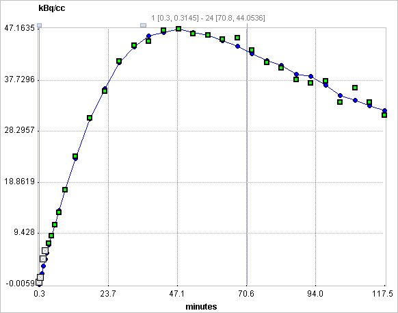
The result of a model fit during Model Preprocessing is shown in the Model Results panel for inspection. The initial points which are not taken into account (before the t* time) are indicated in grey. If no Target tissue is specified, the panel remains empty.

Model Configuration

BPnd |
Binding potential BPnd = k3/k4 . |
k2' |
Clearance rate of the reference tissue. |
-Vt/(Vt'b) |
First multi-linear regression coefficient of the operational equation (Ichise Eq.2.) |
1/b |
Second multi-linear regression coefficient of the operational equation (Ichise Eq.2.) |
-Vt/(Vt'k2'b) |
Third multi-linear regression coefficient of the operational equation (Ichise Eq.2.) |
Reference
1.Ichise M, Liow JS, Lu JQ, Takano A, Model K, Toyama H, Suhara T, Suzuki K, Innis RB, Carson RE: Linearized reference tissue parametric imaging methods: application to [11C]DASB positron emission tomography studies of the serotonin transporter in human brain. J Cereb Blood Flow Metab 2003, 23(9):1096-1112. DOI