An important usage of the VOI analysis is the generation of time-activity curves (TAC) for subsequent kinetic modeling. This can easily be achieved in PVIEW by the following steps
VOI Definition
The image data is loaded as a dynamic series with the correct acquisition times and the correct input units. This is important, because otherwise the acquisition start/end times in kinetic modeling will be wrong, and the TACs may be different in magnitude with respect to the blood data. Such problems result in erroneous model parameters.
In dynamic image series there is generally not enough anatomical information to delineate VOIs. Often, averaging of a subset of the acquisition frames resolves the problem. The VOIs are then delineated in the summed images, transferred to the dynamic images, and optionally saved to a file.
TAC generation
Switch the tool to the dynamic study, and activate the button

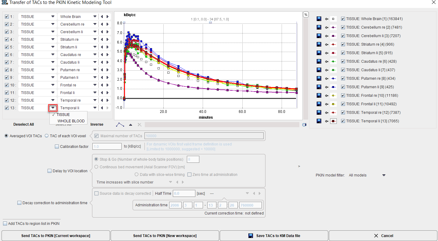
A dialog window appears which is organized in three panels:
1.The left area allows defining the proper type (TISSUE, WHOLE BLOOD) of the calculated TACs and selecting the regions to be sent to the Kinetic modeling tool.
2.The central area shows the TACs.
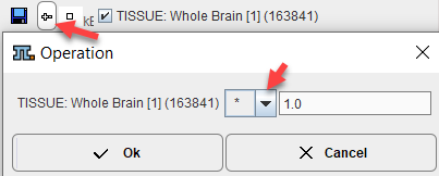
3.The right area controls the displayed TACs. Additionally, the +- buttons allow for simple arithmetic operations with the curve values before transfer to PKIN.
Average TACs
The standard and default procedure is to use the signal average in the VOIs, corresponding to the radio button Averaged VOI TAC. With this setting, not only the average is calculated and transferred, but also the standard deviation which may be used for weighted fits in PKIN.
Voxel-wise TACs
When the transfer mode is set to TAC of each VOI voxel, the individual voxel-wise TACs are transferred. There is a maximum number of TACs which will be considered for the transfer, per default set to 10'000. The number of voxels in the selected VOIs is shown in brackets in the control section. Naturally, the standard deviation is zero in this case.
TAC Value Operations
The calculated TAC values can be modified in two ways.
1.Every individual curve has a +- button which allows multiplying the values with a factor, dividing, adding or taking the logarithm.
2.All generated curves can be scaled with a factor defined by the Calibration factor to kBq/cc option.
TAC Time Shifting
If image data acquisition requires multiple table positions (e.g. dynamic whole-body PET), the timing between VOIs may differ. The timing relative to the injection is crucial for modeling and also for dosimetry. Therefore, the Delay by VOI location option is supported

with two alternatives:
•Stop & Go corresponds to the standard acquisition mode with the table halting in adjacent, overlapping field of views. As there is usually not enough information in the image headers to know for each slice the exact timing, the timing of the different table positions is obtained by dividing the acquisition duration by their number.
•Continuous bed movement assumes that the patient is scanned while the table is continuously moving. Here the axial scanner field of view is required for approximating the time when a VOI is scanned.
Note that the timing derived from these settings can only be approximate and may need correction. The resulting timing of the TAC curves is updated in the curve window whenever one of the settings is changed. Depending on the scanning, the time shifts may be applied in the wrong direction. For instance, the bladder curve may start before the brain curve although the data was acquired in head-in position. To rectify such a case there is a switch Time increases with slice number/Time decreases with slice number.
PKIN Model Filter
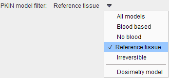
The PKIN model filter selection allows restricting the listed models when the data arrives in PKIN. This useful if the type of data analysis is already known, for instance the modeling with Reference tissue models if no blood data is available. This will also avoid notification messages in PKIN that no blood data is yet available.
With the Dosimetry model selection, the Cumulated Activity model will be set after the TAC data have been transferred to the PKIN tool together with the Total body mass of the patient. This information is extracted from the demographic information in the image, if available.
Decay Correction
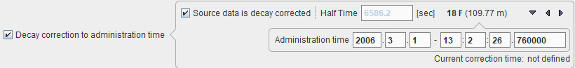
If the Decay correction to administration time flag allows correcting the TACs to an injection time which may be before the first acquisition. If the Source data is decay corrected, only a scaling factor is applied. Otherwise, a time-dependent correction is used.
Data Transfer
The Send TACs to PKIN buttons initiate the transfer of the activity curve data to the PKIN tool. Selecting [Current workspace] transfers the TACs to the currently selected tab in an open PKIN tool. With the Add TACs to region list box checked, the curves are appended as new regions to the existing data, otherwise the current data is over-written. [Current workspace] first creates a new tab in PKIN, to which the data is added. If PKIN is not running, the tool is first started and the data added.
Both the average value and the standard deviation within the VOIs are transferred, as well as patient and study related information. The standard deviation may be used for weighted fits in PKIN.