The timing of the tissue and the blood data should be synchronized to a common clock. Nevertheless, there may be an inherent shift of the blood activity curves relative to the tissue activity curves because of the arrival time difference of the blood in the target tissue and at the sampling location. For brain PET scans with radial artery blood sampling the tracer will appear later in the blood samples than in the brain. This delay is even prolonged if the blood samples are not taken directly with a syringe, but via a catheter which adds additional pathlength as required by online blood sampling devices.
Such a relative delay of the blood samples is accounted for by the Delay parameter of the blood models. Positive delays represent delayed blood information and hence shift the blood curves to earlier times (to the left). While both the whole-blood and the plasma model include a separate Delay parameter, it is reasonable to assume a common delay since the plasma activity is derived from whole-blood. Such a common delay can be fitted together with the kinetic parameters of the tissue model as described below.
Delay Fitting with Single Tissue TAC
The Tissue tab features a dedicated Fit region w. blood delay button. It is hidden underneath the Fit model, current region button and can be activated as illustrated below.
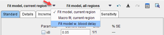
When it is activated, the following actions are performed:
▪The state of the fit flags of the Whole blood and Plasma models is saved.
▪The fit flag of the Delay is enabled, and all other fit flags disabled.
▪A fit of the tissue model is performed, whereby the Delay (common to plasma and whole blood) is also optimized.
▪The state of the fit flags of the Whole blood and Plasma models is restored.
▪The estimated Delay can be inspected below the model parameters, on the Blood tab or in the model history.
The illustration below shows a history of the outcome when fitting a 1- and 2-Tissue compartment model to the WB TAC including the delay.

It is notable that the optimal delay depends on the fitted model, and it will also depend on the selected regional TAC. When using a single TAC it is recommended using the signal from a relatively large VOI, for instance the whole brain, and then keep the delay fixed.
Note: A change of the blood Delay affects the model curve of all regions and therefore requires re-fitting all regional models. If the Delay value is changed manually, it has to be set to the same value for Blood and Plasma.
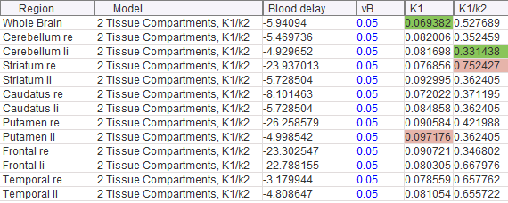
When using Fit model, all regions, the blood parameters will not be fitted separately for each region, even if Float blood parameters is enabled on the Extras panel. Exception is the blood delay: With default Lin. Interpolation model for Blood and Plasma and with the Delay parameter fit enabled, the blood delay will be estimated regionally as illustrated below.
Delay Fitting with Multiple Tissue TACs
An alternative to using a single representative tissue is the use of coupled fitting for the determination of a blood delay which is optimal with respect to tracer appearance in several regions. In the example below 8 regions are selected on the Coupled pane all of which have a 2-tissue compartment model configured. Note that at least one tissue parameters needs to be coupled in this case.
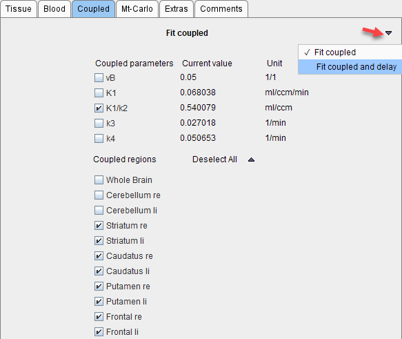
Fitting of the common delay is started using the Fit coupled and delay button which is hidden under the Fit coupled button. The procedure will fit all 8 compartment models while at the same time optimizing the blood timing.