Brain normalization methods are procedures for transforming individual brain images into a standard anatomical coordinate space. They require a suitable template image which is already in the standard space, and an elastic matching procedure for warping the images appropriately. The Brain Normalization (previous) (BN I, Deprecated) and Brain Normalization (formerly BN II) methods have been implemented according to the methodologies used in SPM99 [9] and SPM5, respectively.
Brain Normalization (previous) uses a fixed sampling scheme, whereas Brain Normalization derives the sampling density from the smoothing parameter. Another difference is that Brain Normalization derives the number of basis functions from the specified Frequency cutoff (default = 25) and the Bounding box size. Higher cutoff values result in fewer basis functions.
Note: For work with small animal data only the Brain Normalization method should be applied.
Available Templates (Atlases)
Template (or Atlas) images represent a "standard" anatomy imaged with a certain modality. For the spatial normalization of human brain images the MNI (Montreal Neurological Institute) templates are commonly used. In PFUS, they have been prepared for easy loading as the Reference by the Fusion menu as illustrated below.

It is important to note that the anatomic orientation in the images after loading depends on the menu entry. If a menu entry with HFS (Head First Supine) is selected, the loaded image appears in radiological convention, which is the default for PMOD. For example, loading PET HFS results in the following orientation.

When loading the same images without the HFS arrangement, the result is in HFP (Head First Prone), ie the images are rotated about the z-axis and the face is pointing down as illustrated below.

The available brain templates:
PET / |
PET template provided with SPM5 (Statistical Parametric Mapping). It was constructed by Friston et al. at the Wellcome Department of Cognitive Neurology (University College London, UK) using Oxygen-15 water PET images of 12 normal subjects scanned in resting condition with eyes closed. The template is in MNI (Montreal Neurological Institute) coordinates. |
MR T1 / |
T1 template provided with SPM5. The image was derived from the ICBM152 image which represents the average of 152 healthy T1 brain images by reducing it to 2mm isotropic resolution and smoothing with an 8mm FWHM Gaussian filter. The original ICBM152 data originates from Alan Evans, MNI, Canada (ICBM, NIH P-20 project, Principal Investigator John Mazziotta). The T1 HFS image is also available with the skull removed. |
MR T2 / |
The same as above, but with the T2 MR images. |
User defined |
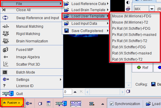
Allows loading a template which is configured in the settings of the PFUS tool as illustrated below.
|

The user can set up additional template images for quick loading with the Load User Template menu entry. Such template images must be in NIfTI format and placed in the Pmod3.6/resources/templates/usertemplates directory. Their proper orientation is up to the user. Note that the menu entry only appears if there are valid templates available.
The PMOD distribution already includes some templates which are described in the PMOD Base Functionality Guide.