If a dynamic series has been loaded and VOIs are defined, the ![]() becomes active, it the PKIN option is included in the PMOD license. At allows calculating the TACs and transferring them directly to the PKIN tool.
becomes active, it the PKIN option is included in the PMOD license. At allows calculating the TACs and transferring them directly to the PKIN tool.
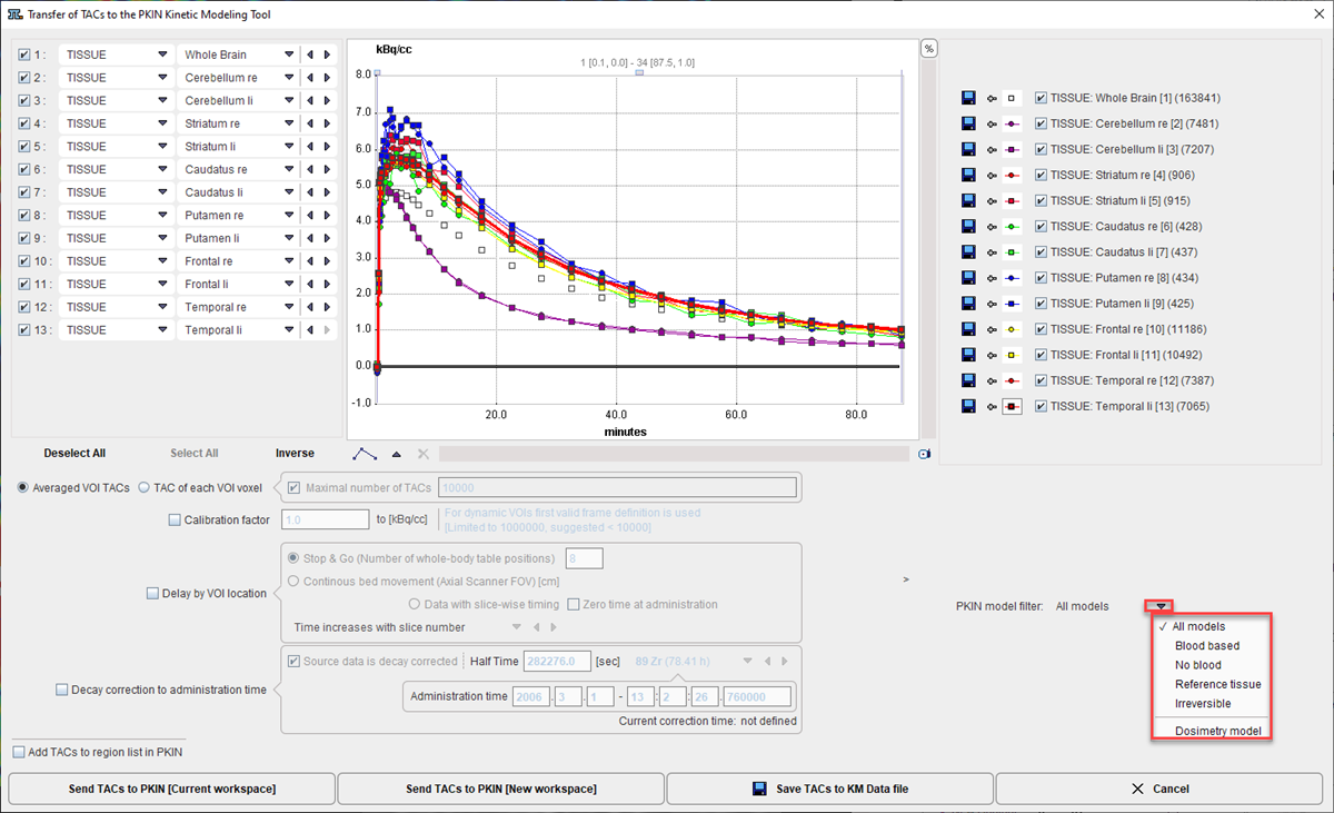
A dialog window appears which is organized in four areas:
1.The left area allows defining the proper type (TISSUE, WHOLE BLOOD) of the calculated TACs and selecting the regions to be transferred.
2.The central area shows the selected TACs.
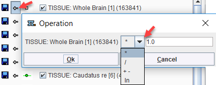
3.The right area controls the displayed TACs. Additionally, the +- buttons allow for simple arithmetic operations with the curve values before transfer to PKIN.
4.The lower area offers several options to modify the calculated TACs.
The following options are supported
Averaged VOI TAC
The standard and default procedure is to transfer the signal average in the VOIs, corresponding to the radio button Averaged VOI TAC. In addition to the average the standard deviation is also calculated and transferred as it is potentially useful for weighted fitting in PKIN.
TACs of each voxel
Instead of transferring the average signal per VOI, the individual TACs of the voxels enclosed in the VOIs can be transferred. This behavior is set by the TAC of each VOI voxel button. For performance reason there is a maximum number of TACs which will be considered for the transfer, per default set to 10'000. The number of voxels in the selected VOIs is shown in brackets in the control section. Naturally, the standard deviation is zero in this case.
TAC Value Operations
The calculated TAC values can be modified in two ways.
1.Every individual curve has a +- button which allows multiplying the values with a factor, dividing, adding or taking the logarithm.
2.All generated curves can be scaled with a factor defined by the Global scale factor to kBq/cc option.
TAC Time Shifting
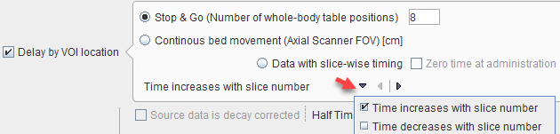
If image data acquisition requires multiple table positions (e.g. dynamic whole-body PET), the timing between VOIs may differ. The timing relative to the injection is crucial for modeling and also for dosimetry. Therefore, the Delay by VOI location option is supported
with three alternatives:
•Stop & Go corresponds to the standard acquisition mode with the table halting in adjacent, overlapping field of views. As there is usually not enough information in the image headers to know for each slice the exact timing, the timing of the different table positions is obtained by dividing the acquisition duration by their number.
•Continuous bed movement assumes that the subject is scanned while the table is continuously moving. Here the axial scanner field of view is required for approximating the time when a VOI is scanned.
•Data with slice-wise timing was introduced to handle data acquired on scanners with continuously moving tables. In this case each slice has an individual time. If the series was acquired after tracer administration (which is recorded in the image), the Zero time at administration box can be enabled to add the time offset to the reference times of the slices.
Note that the timing derived from these settings can only be approximate and may need correction. The resulting timing of the TAC curves is updated in the curve window whenever one of the settings is changed. Depending on the scanning, the time shifts may be applied in the wrong direction. For instance, the bladder curve may start before the brain curve although the data was acquired in head-in position. To rectify such a case there is a switch Time increases with slice number/Time decreases with slice number. For Averaged VOI TAC transfer, the timing of the VOI center will be used.
Model Selection
The Model Selection option allows filtering the list of the PKIN models according to the intended modeling. For the Dosimetry model the Cumulated Activity (OLINDA, IDAC) model will be set after the TAC data have been transferred to the PKIN tool together with the Total body mass of the subject. This information is extracted from the demographic information in the image, and can be edited if it is not present.
Decay Correction
If the Decay correction to Injection time flag allows correcting the TACs to a n injection time which may be before the first acquisition. If the Source data is decay corrected, only a scaling factor is applied. Otherwise, a time-dependent correction is used.
![]()
Data Transfer
The Send buttons initiates the transfer of the activity curve data to the PKIN tool. Selecting Send TACs to PKIN [Current workspace] transfers the TACs to the currently selected tab in an open PKIN tool. With the Append TAC Data box checked, the curves are appended as new regions to the existing data, otherwise the current data is over-written. SendTACs to PKIN [New workspace] first creates a new tab in PKIN, to which the data is added. If PKIN is not running, the tool is first started and the data added. Save TACs to kmData file allows creating a text file which is ready to be used in PKIN batch processing or to process at any later time point in PKIN by loading it through the menu Kinetic/Load KM file.
In addition to the TACs, subject and study related information is also transferred.