Diffusion weighted MR imaging (DWI) has proven a highly valuable tool in brain imaging. It is a quantitative approach allowing for the calculation of various diffusion-related parametric maps. Diffusion tensor imaging (DTI) accumulates even more information and supports the detailed investigation and visualization of fiber tracts where diffusion is highly directional. PMOD has implemented support for DWI and DTI, leveraging the proven DWI/DTI analytics of the CAMINO toolkit [1].
The Diffusion Tensor (DTI MR) model supports the analysis of DWI/DTI data. The results are not only various types of diffusion maps, but also diffusion tensors which can subsequently be used for tractography in PGEM.
Acquisition and Data Requirements
The reading of DWI images with the related gradient information can be a severe challenge. Manufacturers apply proprietary encoding schemes, and research toolkits use their own specific formats for which scientist have developed conversion functions. PXMOD supports DWI data loading in a wide range of formats. Only if at least 6 directions are encoded can the diffusion tensors be calculated. PMOD tries to extract the gradient vectors from the images, if they are DICOM. If this fails or the data are in a different format, the gradient information has to be provided in a text file in preprocessing.
Image Data |
MR data acquired with a DWI sequence. |
Model Preprocessing
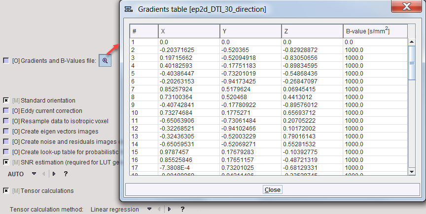
When arriving at model preprocessing, the gradient information should be checked as illustrated below. In this example the directions as well as the gradient size were extracted from DICOM.
If the gradient information is not present, it should be prepared in one of the supported formats and then configured as illustrated below.
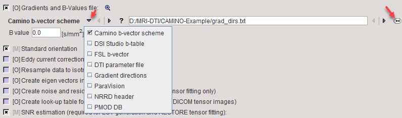
The ? button to the right provides a description of the supported gradient text formats which is reproduced below. B-balues have to be specified in [s/m2].
Camino b-vector scheme |
This text file contains four columns of data. The first three columns are the b-vectors for x, y, and z. The fourth column is for the b-value. |
DSI Studio b-table |
This text file contains four columns of data. The first column is for the b-value. The following columns are the b-vectors for x, y, and z. |
FSL b-vector |
This text file contains the b-vectors for x, y, and z in three rows. As PMOD does not support b-value files as a separate input you may add the b-values as a fourth row. |
DTI parameter file |
This option retrieves b-vectors and b-values from a DTI Studio parameter file. |
Gradient directions |
This general option will accept text files with three to five columns. The first three columns are the b-vectors for x, y, and z. A four-columns file is assumed to have the b-values in the last column. A five-columns file is assumed to have gradient indexes in the first column. This option accepts gradient tables exported from the Pmod Info Dialog. |
ParaVision |
This option retrieves b-vectors and b-values from the ParaVision Parameter List file. |
NRRD header |
This option retrieves b-vectors and b-values from the DWMRI section of the NRRD formatheader file. |
PMOD DB |
This option retrieves b-vectors and b-values from a gradient table file stored to PMOD database. The data in the file is arranged in five columns as in Gradient directions file. Comment lines start with #. |
Several options are available
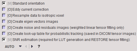
Eddy current correction |
Correction for eddy-current-distortions and motion artifacts, based on the matching procedure with averaged and motion-corrected b0 image. |
Resample data to isotropic voxel |
Resampling the data in time domain in order to obtain volume data with uniform timing. |
Create eigen vector images |
Rigid matching relative to first frame. |
Create noise and residuals images |
These images can only be produced by the Weighted linear tensor fitting. |
Create look-up table for probabilistic tracking |
This information is required for probabilistic mapping using the PICo mehtod in PGEM. It is saved with the tensor images using private DICOM fields. |
SNR estimation |
Noise estimation is required for initialization of the RESTORE tensor calculation method. PMOD supports the traditional background ROI approach and two methods based on multiple B0 images in the DWI series, all as implemented in UCL Camino Diffusion MRI Toolkit. The multiple B0 images methods are preferred over background ROI method. AUTO: Background (noise) ROI or signal (white matter) ROI will be estimated as needed based on three levels thresholding. If a mask is defined it will be used to define the background ROI. Depending on the number of B0 images in the series one of the supported noise estimation methods will be applied. RATIO: Provided sigma value will be used as an estimation of the noise standard deviation in the image. VOI: Provided ROI will define the signal region for estimation methods based on multiple B0 images. The estimation method used will be still determined by the number of B0 images so in case of single B0 image that configuration has no effect and the result will be the same as in the AUTO option. |
Four different calculations can be applied for tensor calculation, which invoke the Camino modelfit function.
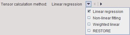
Linear |
Uses unweighted linear least-squares to fit the diffusion tensor to the log measurements [2]. |
Non-linear |
Provides more accurate noise modeling than linear fitting [3]. |
Weighted linear |
Uses weighted linear least-squares fitting [3]. |
RESTORE |
Fits the diffusion tensor robustly in the presence of outliers [4]. |
Model Configuration
Images of the following parameters can be generated
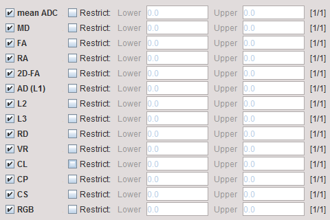
mean ADC |
Mean apparent diffusion coefficient. |
MD |
Mean diffusivity. Characterizing the net degree of displacement of the water molecules. |
FA |
Fractional anisotropy. A ratio ranging from 0 to 1 that represents the degree to which diffusion is anisotropic. High FA values indicate that diffusion is much greater in one direction that others, whereas low FA values indicate that diffusion is nearly equal in every direction. |
RA |
Relative anisotropy, also a measure of diffusion anisotropy. |
2D-FA |
Two-dimensional fractional anisotropy. |
AD(L1) |
Axial (or longitudinal) diffusivity. Rate of diffusion in the principal diffusion direction, i.e. the first eigenvalue. |
L2 |
Second eigenvalue. |
L3 |
Third eigenvalue. |
RD |
Radial diffusivity. Rate of diffusion perpendicular to the principal diffusion direction. |
VR |
Volume ratio, another measure of diffusion anisotropy. |
CL |
Tensor ellipsoid linearity. |
CL |
Tensor ellipsoid planarity. |
CS |
Tensor ellipsoid sphericity. |
RGB |
Direction of the principal diffusion direction encoded as an RGB image. Red indicates diffusion along the x-axis, left-right orientation; green indicates diffusion along the y-axis, posterior-anterior orientation; and blue indicates diffusion along the z-axis, inferior-superior orientation. The map provides an indication of whether the white matter tracts are in proper orientation. |
However, pixelwise calculation adds additional results which can be found in the list of parametric maps.
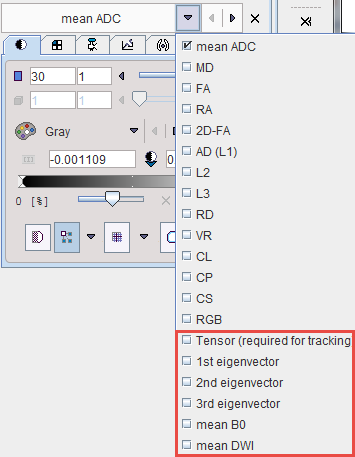
Tensor |
This is the input dataset for tractography. It is encoded as a dynamic series with 6 dimensions to encode all tensor information. |
1st eigenvector |
These data sets contain the tensor eigenvectors which are encoded in a dynamic series with 3 dimensions. |
residuals |
Residuals after fitting the tensor. |
mean B0 |
Average of all images in the DWI series without diffusion gradient. |
mean DWI |
Average of all images in the DWI series with diffusion gradient. |
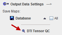
Parametric Maps
Note that on the Parametric Maps page there is a button DTI Tensor QC.
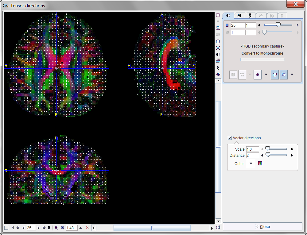
When activated it shows a window for controlling the quality of the results. It shows the projection of the principal diffusion direction vectors together with the color-coded direction image. Red indicates diffusion along the x-axis (left-right), green diffusion along the y-axis (posterior-anterior), and blue diffusion along the z-axis (inferior-superior).
References:
See also Camino References.
1.Cook P. A., Bai Y., Nedjati-Gilani S., Seunarine K. K., Hall M. G., Parker G. J., Alexander D. C., "Camino: Open-Source Diffusion-MRI Reconstruction and Processing", 14th Scientific Meeting of the International Society for Magnetic Resonance in Medicine, Seattle, WA, USA, p. 2759, May 2006.
2.Basser P.J., Mattielo J., and Lebihan D., "Estimation of the effective self-diffusion tensor from the NMR spin echo, Journal of Magnetic Resonance", 103, 247-54, 1994.
3.Jones D.K. and Basser P.J., "Squashing peanuts and smashing pumpkins: How noise distorts diffusion-weighted MR data", Magnetic Resonance in Medicine, 52(5), 979-993, 2004.
4.Chang L-C, Jones D.K. and Pierpaoli C., "RESTORE: Robust estimation of tensors by outlier rejection, Magnetic Resonance in Medicine", 53(5), 1088-1095, 2005.