The result of matching is shown on the MATCHED PET page. Please verify that matching was successful by evaluating the alignment in different parts of the brain. Particularly helpful to do so is to interactively drag the fusion balance left/right, and to enable contour outlines.
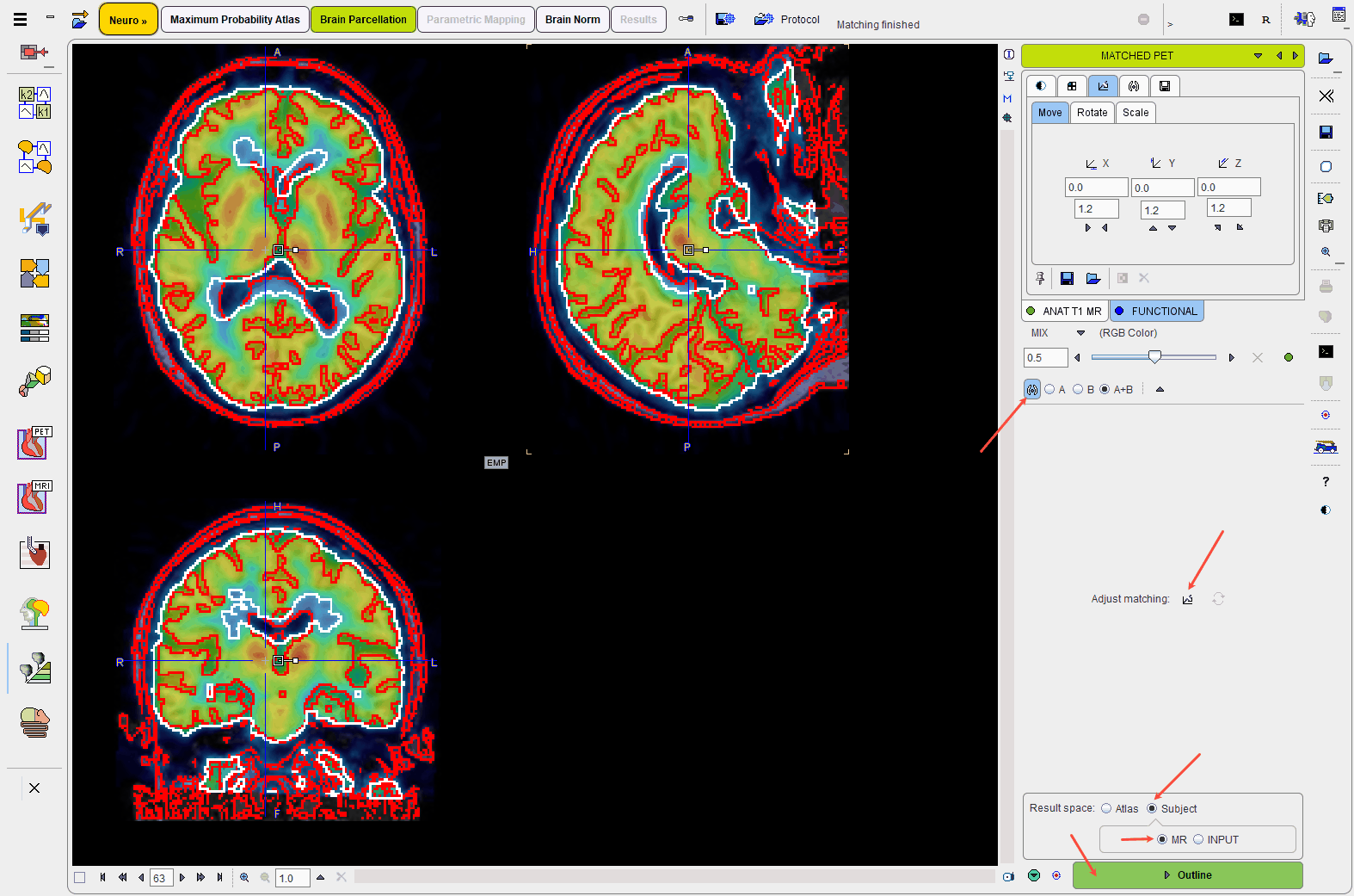
If the match is not satisfactory, there are two options to rectify the situation:
1.Return to the previous page, change the sampling and smoothing parameters and try the automatic matching again, or
2.Activate the Adjust matching button and shift/rotate the PET image interactively by dragging the handles in the image or entering offsets/angles on the Move/Rotate tabs.
Result Space
The next step consists of outlining the brain segments in the Result space which defines, where the PET statistics are calculated:
Atlas |
The PET image is transformed to the Atlas space and the statistics are calculated with interpolated PET values. |
MR |
The PET image is transformed to the MR space and statistics are calculated with interpolated PET values. |
PET |
The brain structures are mapped to the PET space and statistics are calculated with the original PET values. |
Outlining of the brain structures in the Result space is started with the Outline button.