The Retention Fraction is the fraction of the total tracer delivered to an organ that is extracted into and retained by the tissue. It is the residual after clearances of the vascular component and the portion of the tracer that rapidly back diffuses from tissue to blood. Usually this fraction of tracer is sequestered in more slowly turning over metabolic or membrane binding processes. [1]
Operational Equation
Given a tissue TAC CT(t) and the input curve CP(t) this auxiliary model allows calculating the retention fraction R by [2]:

The denominator with the input curve integrated from the time of injection represents the available tracer, the numerator with the integral of the late tissue uptake the actually extracted tracer. Other authors [3] have used a simplified version of tracer retention which is calculated by dividing the tracer concentration by the input curve integral.
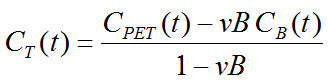
Note that an explicit blood volume correction is available via the vB input parameter. In the case of vB=0 (default setting), no correction is performed. For vB>0, the tissue TAC is corrected by the scaled whole blood activity before the actual analysis as follows.
Implementation
The Tracer Retention auxiliary model has two input parameters Start and End for the specification of tb and te in the integration formula above, respectively. The curve display shows the input curve interpolated at the frame mid-times. The result parameters are calculated in % and %/min for the two calculation methods:
R_window |
Retention of tracer in the time window (Tissue-Integral in window)/(Plasma-Integral from start) |
R_wind_min |
Retention of tracer in the time window (Tissue-Average in window)/(Plasma-Integral from start) |
R_last |
Retention of tracer in the last frame of the acquisition (Tissue-Integral in window)/(Plasma-Integral from start) |
R_last_min |
Retention of tracer in the last frame of the acquisition (Tissue-Average in window)/(Plasma-Integral from start) |
References
1.see Glossary in Phelps ME: PET : molecular imaging and its biological applications. New York: Springer; 2004.
2.Hutchins GD, Chen T, Carlson KA, Fain RL, Winkle W, Vavrek T, Mock BH, Zipes DP: PET imaging of oxidative metabolism abnormalities in sympathetically denervated canine myocardium. J Nucl Med 1999, 40(5):846-853.
3.Di Carli MF, Tobes MC, Mangner T, Levine AB, Muzik O, Chakroborty P, Levine TB: Effects of cardiac sympathetic innervation on coronary blood flow. The New England journal of medicine 1997, 336(17):1208-1215.