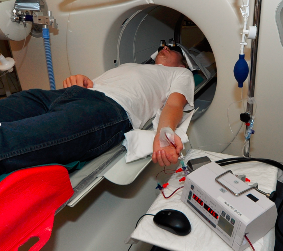
Automated Blood Sampling Setup
Automated blood sampling is the preferred method for measuring the radioactivity concentration in whole blood. In such a setup, a catheter is inserted into an artery, and blood continuously run through a detector which measures the radioactivity from the time of tracer injection until the end of the PET acquisition. If needed, automated blood sampling can be stopped when the blood concentration changes have become slow, and some manual blood samples be withdrawn at later times.
A typical blood sampling setup is illustrated below with a catheter in the radial artery, which is run through a coincidence detector (twilite, www.swisstrace.com) and a pump which ensures constant blood drawing speed. Three-way valves serve for occasionally taking blood assays, which are needed for measuring the metabolite build-up over time.
Blood Dispersion
Because blood needs to travel an extra distance outside the body until it reaches the detector, the blood measurements are delayed. Furthermore, the time course of the concentration is altered, because fast concentration changes are smoothed in the catheter, an effect called "dispersion". In kinetic modeling, results can be biased if dispersed blood information is used, particularly for short measurements such as 15O-water bolus studies for quantifying tissue perfusion.
The sections below describe the methodology and implementation of a dispersion correction methodology developed by Munk et al. [1].