The Card NH3 (2-Tissue) model developed by Hutchins et al. [1] is an implementation of the irreversible 2-tissue-compartment model for cardiac PET studies using 13NH3 ammonia bolus injection. The compartment model has the following structure
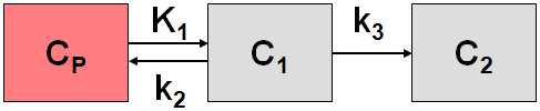
where C1 is free tracer in tissue, and C2 is metabolically trapped tracer in the form of 13N glutamine. Because ammonia is considered in this model as freely diffusible across the capillary wall, the unidirectional uptake parameter K1 equals the myocardial perfusion.
Operational Model Curve
The system of differential equations is

To allow the fitting of data over an extended period, the model includes the exponential metabolite correction described by van den Hoff et al. [2]

with a delay t0=0.48 min and half-time T1/2=6.69 min. CLV(t) is the total tracer concentration measured in the left ventricular cavity, including metabolites.
Additionally, the model incorporates a cardiac dual spillover correction by the operational equation
![]()
where
VLV = spill-over fraction of the blood activity in the left ventricle CLV(t),
VRV = spill-over fraction of the blood activity in the right ventricle CRV(t) .
Implementation Notes:
When using the model from the PCARD module, the data are transferred appropriately. When using it in PKIN the blood data have to be loaded as follows:
▪The left ventricle curve must be loaded as the Plasma activity curve. No metabolite correction needs to be enabled on the Blood panel of PKIN because it is included in the tissue model.
▪The right ventricle curve must be loaded as the Whole blood curve.
The following automatic adjustments are performed within the model:
▪The spill-over fraction from the right ventricle VRV is automatically fixed to zero if the string "Sep" is not contained in the name a region. The assumption is that such a TAC is not from septal tissue and should thus be modeled with spill-over from the left ventricle only.
▪The valid flag for all data samples after 2 minutes is set to false, and they are consequently not considered in the fit.
Parameter Fitting
The model includes the five fitable parameters F, vLV, vRV, k2, k3. Please inspect the %SE standard error to get information about the reliability of their estimates. The input parameters MC T0 (0.48) and MC T12 (6.69) are related to the metabolite correction as described above and can be adjusted if needed.
Alternate Model Card NH3 (2-Tissue, K1/k2)
Due to the increased number of fit parameters it has been found, that the Card NH3 (2-Tissue) may suffer from identifiability problems. Therefore, the variant Card NH3 (2-Tissue, K1/k2) has been developed using the parameter K1/k2 (distribution volume of the first compartment) as a fit parameter instead of k2. In this configuration physiological restrictions can be imposed on K1/k2 , or K1/k2 can be used as a common parameter in a coupled fit.
References
1.Hutchins GD, Schwaiger M, Rosenspire KC, Krivokapich J, Schelbert H, Kuhl DE: Noninvasive quantification of regional blood flow in the human heart using N-13 ammonia and dynamic positron emission tomographic imaging. J Am Coll Cardiol 1990, 15(5):1032-1042.
2.van den Hoff J, Burchert W, Borner AR, Fricke H, Kuhnel G, Meyer GJ, Otto D, Weckesser E, Wolpers HG, Knapp WH: [1-(11)C]Acetate as a quantitative perfusion tracer in myocardial PET. J Nucl Med 2001, 42(8):1174-1182. PDF