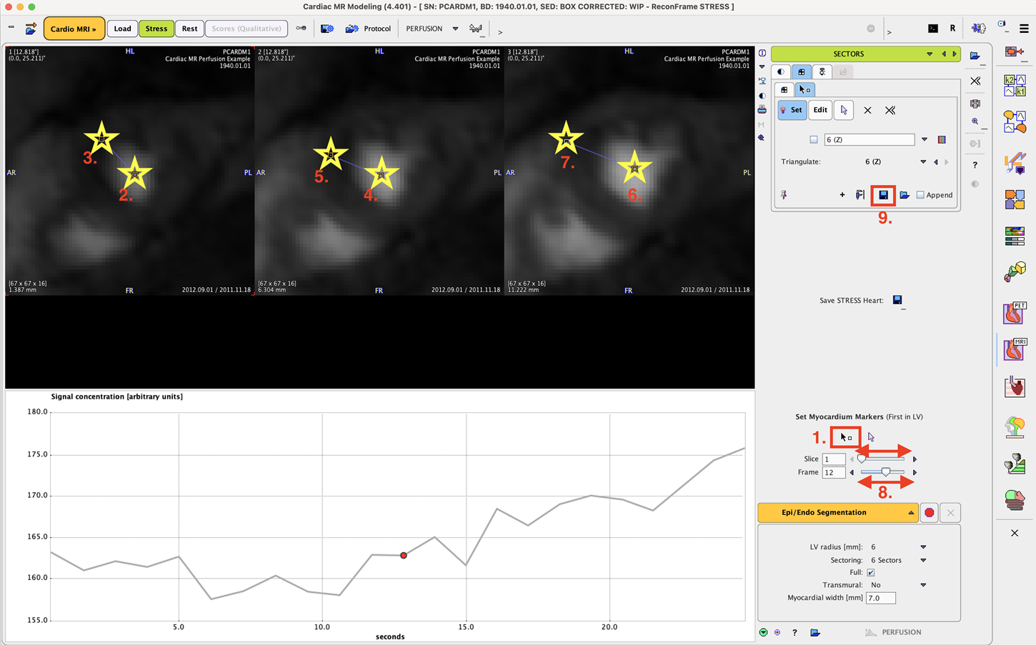
The first step consists of defining in each relevant slice the center of the LV cavity and the edge of the right ventricle.

Please proceed as described below and indicated in the illustration above by the numbers.
1.Switch on markers definition by activating the ![]() button.
button.
2.In each slice, first click into the LV center, then to the RV edge as illustrated.
3.Use the Slice slider to move to the next short-axis slices. Repeat markers placement, until all slices are covered.
4.Use the Frame slider to change the image contrast.
5.Optionally, save the markers definition with the indicated button (9.). Note that the markers are automatically saved if the data resides in a PMOD database.