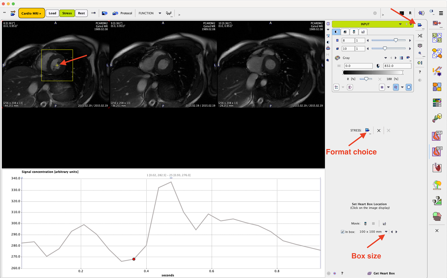
For the analysis of cardiac function, cine MR images in short-axis orientation are required, ideally with the right ventricle located to the left of the images. Note that the images can't be reoriented during the workflow, so that reorientations have to be performed before loading in PCARDM.
Image Loading
The images can be loaded from different locations, via the Load DATABASE page, the loading button in the taskbar to the right, or with the Load button on the INPUT page. The two tabs Stress and Rest incorporate the same workflows for two data sets, and their results can be compare in a dialog window.

Image Cropping
For the automatic segmentation of the ventricle the cropping of the volume around the heart might be helpful. To this end, the In box field can be checked and the box size changed with the arrow button next to it. The location of the box is changed by simply clicking into the center of the ventricle in any of the images at the top. It is recommended to position the image at a basal slide in diastole before placing the box. Note that the volume is only cropped in the x and y dimension, whereas all slices will be retained.
The button Get Heart in Box initiates cropping and proceeds to the next page SEGMENTS.