The data should be cropped in two ways:
1.spatially, to remove signal remote to the heart, and
2.in the time domain, to only include proper first-pass signal.
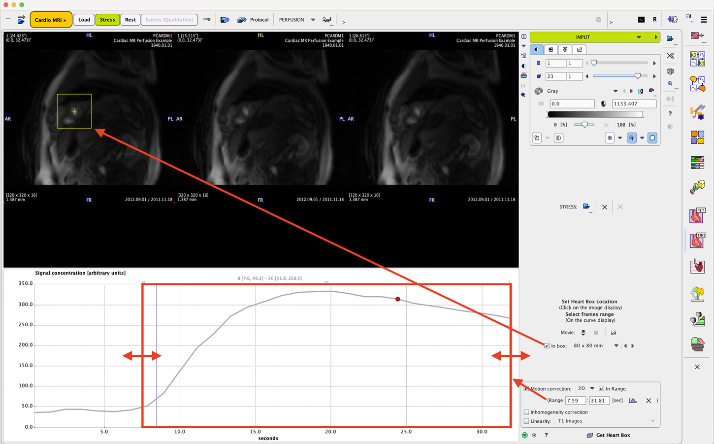
Heart Cropping
If the In box is checked, a square is shown in the images which should be centered on the left ventricle. To do so, just click into the center of the cavity. Verify that all slices of the LV are within the box by moving the slices slider left/right. If the box is too tight, a bigger size can be selected from the size list. Note that the motion of the heart during the acquisition can be inspected by moving the times slider. Ideally, motion due to respiration is only minimal.
Time Range
The time range of the data used for the perfusion quantification should be restricted to the first-pass phase. The initial part is removed to avoid a signal bump or other erroneous signal contributions specifically for kt-PCA reconstructed data due to the temporal correlations included in the reconstruction process. It is particularly important to avoid late signal with recirculating contrast agent. The start and end times can be specified numerically in the Time range section. Alternatively, corresponding markers can be dragged directly in the curve area as indicated above.