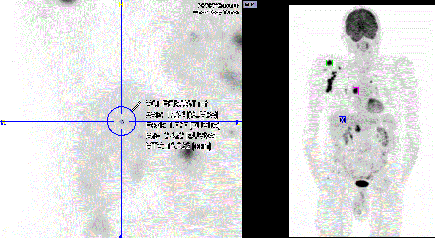
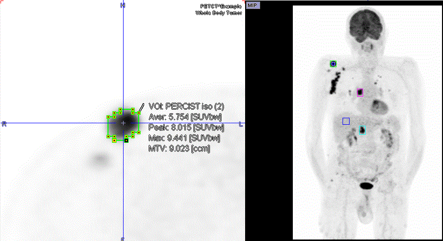
Instead of basing iso-contouring on the values in the lesion itself or an absolute SUV, the threshold can be obtained from the uptake in reference tissue. The PERCIST (PET Response Criteria in Solid Tumors) [1,2] obtains the reference activity from a 3cm diameter sphere placed in the right side of the liver, midway between the dome and inferior margin, excluding central ducts and vessels. If the liver is diseased, background is to be measured in the descending thoracic aorta (cylinder: 1cm diameter, 2cm long, avoiding wall).
The procedure for a PERCIST-conformant assessment using hot-key based VOis is as follows:
1.Activate the VOI functionality.
2.Reference sphere: Triangulate the point in the liver as described above. Use keyboard shortcut Ctrl+Shift+U to place the 3cm diameter reference sphere. An entry VOI named PERCIST ref appears in the VOIs list. The minimal level of tumor uptake is calculated as 1.5*AverageLiver+2*StandardDeviationLiver. This value is used as iso-contouring threshold in the subsequent lesion assessment.

2.Lesions: For each of the lesions triangulate its center, and then use keys Ctrl+U. A region growing algorithm is applied and the lesion outlined on the threshold level calculated from PERCIST ref. If the lesion is below the minimal tumor level, no iso-contouring VOI is found. In this case the lesion is not measurable with PERCIST. Otherwise a VOI is enteredd using

For each measurable lesion a corresponding entry will appear in the VOIs list named PERCIST iso followed by a number through round brackets, e.g. (3), indicating the order of the outlining.
VOI Sorting can be used for bringing the most relevant lesions to the top. For individual lesion documentation it is recommended to enable Overlay Statistics and then click at the VOIs in the list and perform an Image Capture with Ctrl+E.
References
1.Wahl RL, Jacene H, Kasamon Y, Lodge MA: From RECIST to PERCIST: Evolving Considerations for PET response criteria in solid tumors. J Nucl Med 2009, 50 Suppl 1:122S-150S.
2.O JH, Lodge MA, Wahl RL: Practical PERCIST: A Simplified Guide to PET Response Criteria in Solid Tumors 1.0. Radiology 2016, 280(2):576-584