There are various approaches for outlining a lesion in a PET image. The PET Tumor volume segmentation method implements an automatic model-based approach [1, 2] for determining an iso-contouring threshold which is independent of the signal-to-background ratio and provides accurate volume measurements down to the resolution of the PET scanner.
Principle
The method performs an iterative adjustment of the iso-contouring level, taking into account the scanner resolution and the background activity. In this process, the lesion geometry is approximated by a sphere and the appropriate threshold level determined by a lookup procedure which refers to a homogeneous sphere imaged at scanner resolution. During the iterations, each threshold setting results in a new tumor volume obtained by iso-contouring, which allows calculating the next threshold approximation. The process is repeated until the tumor volume becomes stable.
Required for applying this segmentation method is:
1.An initial approximation of the tumor VOI.
2.The background activity level which is subtracted.
3.The PET scanner resolution.
Use without Background VOI
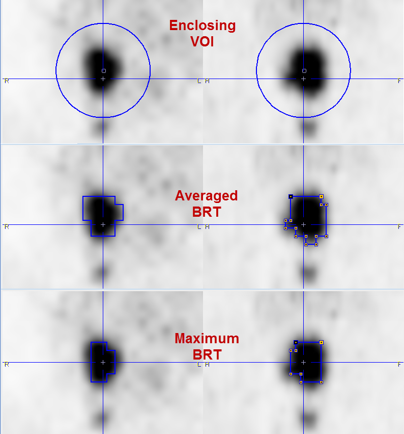
If the tumor is isolated from the surroundings, the background can easily be obtained from the initial VOI definition. Please define a VOI enclosing the tumor, with sufficient distance to the rising edge. A sphere is used in the illustration below.
Start the Tumor Volume Segmentation procedure. In the appearing dialog window
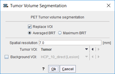
enter the Spatial resolution of the image. There are two different approaches implemented. With Averaged BRT (Boundary-Reproducing Threshold) the average activity in the iteratively segmented VOI is used [1], with Maximum BRT the maximal activity [2]. According to Jentzen [1], the Averaged BRT is preferable for larger objects, and it is less sensitive to noise and non-uniform uptake.
Check Replace VOI, if the initial VOI is not used any further and start the iteration with Ok. The program first calculates the background activity by dilating the bounding VOI by 3 pixels and averaging the pixels along the extended contour. Then the optimal iso-contouring threshold is calculated as outlined above. The results obtained with the different criteria are illustrated below.
Use with Background VOI
If higher uptake than background is close to the tumor, the initial VOI should be close to the tumor boundaries, and an explicit background VOI should additionally be outlined. Note that the Background VOI box needs to be checked, and the proper VOI selected from the list next to it.
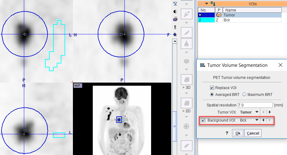
Note: The Tumor Volume Segmentation is called while the Group tab is active, all selected VOIs will be processed. If a background VOI is defined and named "Bck" it will be automatically considered by the algorithm.
References
1.Jentzen W.: An improved iterative thresholding method to delineate PET volumes using the delineation-averaged signal instead of the enclosed maximum signal. Journal of nuclear medicine technology 2015, 43(1):28-35. DOI
2.van Dalen JA, Hoffmann AL, Dicken V, Vogel WV, Wiering B, Ruers TJ, Karssemeijer N, Oyen WJ: A novel iterative method for lesion delineation and volumetric quantification with FDG PET. Nuclear medicine communications 2007, 28(6):485-493. DOI