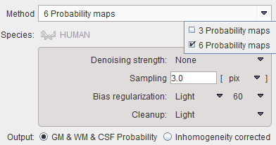
The MRI Probability/Inhomogeneity tool allows applying the unified segmentation methods of SPM8 or SPM12 to the loaded T1-MR image. Depending on the Output setting, the Grey Matter, White Matter, CSF probability maps are saved as frames in a dynamic series, or the MR image corrected for the spatial inhomogeneity.

Method |
Two SPM-type segmentation variants are supported, the 3 Probability Maps (SPM8) and the 6 Probability Maps (SP12) variant. Note that the normalization transform which is obtained as part of the segmentation can be applied for the spatial normalization of the subject brain anatomy in later stages. |
Denoising strength |
Denoising of the MR image may improve the segmentation of gray matter, white matter and CSF. If a Denoising strength other than None is selected, a non-local means denoising algorithm is applied which preserves structure boundaries unless the strength is too high. |
Sampling |
Density of pixels considered in the calculation. It can be specified in pixel or mm units. |
Bias regularization |
Serves for compensating modulations of the image intensity across the field-of-view. Depending on the degree of the modulation, a corresponding setting can be selected from the list. The parameter to the right indicates the FWHM [mm] to be applied. The larger the FWHM, the smoother the variation is assumed. |
Cleanup |
Procedure for rectifying the segmentation along the boundaries. |
It is recommended to use the default settings and only experiment with other parameter values if the segmentation fails. The default settings can be recovered by the ![]() button.
button.